Download
1 / 46
500 likes | 1.13k Vues
Pathology 6 White blood cell and lymph node disorders (1). Toxic changes in neutrophils include all the following, except: Cytoplasmic basophilia Large basophilic granules Hypersegmentation Cytoplasmic vacuoles Dohle bodies. Large granular lymphoma is associated with Neutropenia

E N D
Toxic changes in neutrophils include all the following, except: • Cytoplasmic basophilia • Large basophilic granules • Hypersegmentation • Cytoplasmic vacuoles • Dohle bodies
Large granular lymphoma is associated with • Neutropenia • Lymphocytopenia • Neutrophilia • Eosinophilia • Basophilia
Monocyopenia is associated with which one of the following tumors • Lymphoplasmacytic lymphoma • Chronic lymphocytic leukemia • Large granular lymphoma • Hairy cell leukemia • Hodgkin lymphoma
Nonneoplastic WBC disorders. • High number (cytosis) • Low number (cytopenia) • Abnormal function
5 major types of WBCs • Neutrophils • Lymphocytes • Monocytes • Eosinophils • Basophils
What is the characteristic finding that lymphocytes display on peripheral blood in patients with pertussis? • Reactive lymphocytes • Lymphocytes with cleaved nuclei • Normal lymphocytes • Prominent nucleoli • Hairy cytoplasmic extensions
Neutropenia • Decrease in the number of neutrophils • When severe, it is called agranulocytosis • Usually less than 500 • Increased risk of infection • Two major mechanisms • Decrease production • Increase peripheral destruction of neutrophils
Decrease production • Marrow hypoplasia in patients who receive chemotherapy • Leukemia or other tumors replacing the marrow • Medications • Certain types of neoplastic lymphocytic proliferations such as large granular leukemia (LGL)
Increased peripheral use • Autoimmune destruction • Overwhelming bacterial, fungal or rickettsial infection • splenomegaly
Clinical manifestations • Infection, infection, infection!!! • bacterial • Fever, chills, malaise • Mucocutaneous necrotizing ulcers • High risk of sepsis
Treat with broad spectrum antibiotics • Depending on the clinical setting, treat with G-CSF
lymphocytopenia • Low number of lymphocytes • Less than 1000 in adults • Less than 3000 in children <2 years of age
Increased risk of • Opportunistic infections • Autoimmune disorders • Malignancy
Causes • Acquired: • HIV/AIDS • Protein malnutrition • Viral infections • Autoimmune disorders: SLE and RA • Certain leukemia or lymphoma tumors
Inherited • Severe combined immunodeficiency • Wiscott-Aldrich syndrome
Clinical manifestations • Infections with unusual microoganisms • Pneumocystis jiroveci • Fungal infections • CMV • Zoster These infection could be fatal • Risk of malignancies • Autoimmune disorders
Symptoms of associated diseases • Small or absent tonsils or lymph nodes in HIV or immune deficiency diseases • Lymphadenopathy: again in early HIV or lymphoma • Eczema in WAS • Stigmata of pancytopenia in cases of leukemia
Treatment • Treat infections • Treat underlying condition • IVIG for certain types of immune deficiency • Bone marrow transplantation
Monocytopenia • In the setting of other pancytopenia • Associated with hairy cell leukemia
eosinopenia • Can be found in healthy individuals • Of limited clinical significance • Steroid use • Acute inflammatory condition
Basopenia • Limited clinical significance • Acute hypersensitivity • Thyrotoxicosis • Infection
Neutrophilia • Increased number of neutrophils • Caused by • Acute bacterial infections • Sterile inflammation • Burns • Myocardial infarction • Morphology: TOXIC CHANGES
Toxic changes • Occur in the setting of bacterial infection • Cytoplasmic basophilia • Vacuoles • Dohle bodies
Lymphocytosis • Increased number of lymphocytes • Chronic bacterial infections • TB, brucella • Pertussis • Viral infection • Morphology depends on the cause
Infectious mononucleosis • Reactive lymphocytes
Monocytosis • Increased number of monocytes • Chronic infections such as TB (associated with lymphocytosis) • Endocarditis • Rickettsial infection • Collagen vascular disease such as SLE • Inflammatory bowel disease • Certain myeloid leukemia with monocytic differentiation
Eosinophilia • Allergic disorders • Parasitic infections • Medications • Collagen vascular disorders • Vasculitis • Atheroembolic disorders (transient) • Certain myelproliferative neoplasms • Certain lymphoma, specifically Hodgkin lymphoma
Basophilia • Rare, • Usually in the setting of chronic myelogenous leukemia.
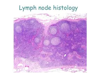
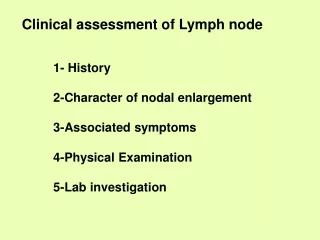
Acute nonspecific lymphadenitis • Enlargement of a localized or generalized group of lymph nodes • Localized in cases of draining a site of infection • Generalized viral or bacterial infection
Painful enlargement of the lymph nodes. • Histology: • Large follicles with germinal center formation • Frequent GC macrophages • Sinus enlargement with histiocytes • Parafollicular neutrophils, necrosis and possible pus formation
Chronic Nonspecific Lymphadenitis • It assumes three patterns • Follicular hyperplasia • Paracortical hyperplasia • Sinus histiocytosis
No neutrophils • Can be confused with follicular lymphoma (1) the preservation of the lymph node architecture; (2) variation in the shape and size of the germinal centers; (3) the presence of a mixture of germinal center lymphocytes of varying shape and size; (4) prominent phagocytic and mitotic activity in germinal centers. (5) BCL2 is negative in the follicles and positive in follicular lymphoma
Paracortical hyperplasia • Viral infections • Medications • After vaccines
Toxic changes in neutrophils include all the following, except: • Cytoplasmic basophilia • Large basophilic granules • Hypersegmentation • Cytoplasmic vacuoles • Dohle bodies
Large granular lymphoma is associated with • Neutropenia • Lymphocytopenia • Neutrophilia • Eosinophilia • Basophilia
Monocyopenia is associated with which one of the following tumors • Lymphoplasmacytic lymphoma • Chronic lymphocytic leukemia • Large granular lymphoma • Hairy cell leukemia • Hodgkin lymphoma
What is the characteristic finding that lymphocytes display on peripheral blood in patients with pertussis? • Reactive lymphocytes • Lymphocytes with cleaved nuclei • Normal lymphocytes • Prominent nucleoli • Hairy cytoplasmic extensions