Download
1 / 9
0 likes | 6 Vues
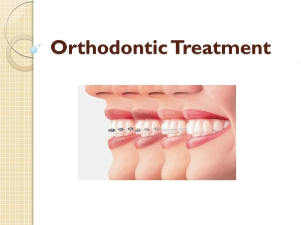
Accelerated Orthodontic Tooth Movements

E N D
1 Accelerated Orthodontic Tooth M Accelerated Orthodontic Tooth Movement ovements s Prepared by: Prepared by: Dr Mohammed Alruby Dr Mohammed Alruby الله نم بلطا نكلو هلهس هايح كيطعي نا الله نم بلطت لا هبعصلا هايحلا ةهجاوم يلع هوقلا كيطعي نا Accelerated orthodontic Tooth Movements Accelerated orthodontic Tooth Movements Dr. Mohammed Alruby Dr. Mohammed Alruby
2 Accelerated tooth movement in orthodontic is a challenging task to shorten the treatment time Research in this area confined into the following categories; 1-Biomechanical approach: as self-ligating system 2-Physiological approach: such as direct electric stimuli, or low level Laser therapies (LLLTs) 3-Pharmacological approach: local injection of cytokines or hormones 4-Surgical assisted approach: periodontal ligament distraction, dento-alveolar distraction, selective decortication, 5-Surgery simulated approach: as submucosal injection of platelets rich plasma (PRP) 1-Biomechanical approach: self-ligating bracket system = 1st self-ligating -------- Russell attachment 1935 = edge lock (oramco) ----- 1972 = mobile lock (Forstadent) ------1980 = speed ---------------- 1980 = active --------------- 1986 Self-ligating brackets has 2 categories, active and passive Active: bracket have a spring clip that store energy to pass against the arch wire Passive: bracket have slide that can be closed and does not encroach on slot lumen Self-ligating bracket enable tooth to slide along an arch wire with lower and more predictable net forces with complete control Mechanism: The primary advantage of self-ligating over conventional that occurs because the usual steel or elastomeric ligature not necessary Passive design generates less friction than active one. Under conventional, the friction / bracket with Niti wire was 41gm in Dentaurum bracket, and 15gm with Damon bracket with stainless steel wire With reduced friction may become 3.6gm so less force needed to produce movement Self-ligating bracket produce more physiologically harmonies tooth movement by interrupting periodontal vascular supply so: -More alveolar bone generation -Greater amount of expansion -Less Proclination of anterior segment -Less need for extraction ** several systematic reviews and studies revealed that self-ligating bracket do not accelerate alignment or space closure in clinical setting, this approach paradox in likely due to the effect of binding because when the teeth tip, rotate or torque, the edges of slot engage the arch wire creating binding so that resistance to sliding increase. ** because the bracket design of self-ligating is narrower than conventional type so the effect of binding is greater resulting in increased resistance to sliding compared with conventional. Less incisor Proclination appear the more advantage of self-ligating bracket ** tooth movement is a metabolic process of alveolar bone resorption and deposition of bone, so acceleration of movement may affect by biological and surgical procedure 2-Physiological approach: direct electric current stimulation: Beason et al, the 1st that proposed use of electric current for orthodontic tooth movement near to tooth that moved but failed to demonstrate the effect on movement Accelerated orthodontic Tooth Movements Accelerated orthodontic Tooth Movements Dr. Mohammed Alruby Dr. Mohammed Alruby
3 DavidoVitch et al reported successful results in accelerating orthodontic tooth movement through direct current on gingival tissue as near as possible to the moving teeth Mechanism: Direct electric current was 7 volt and 15 microamperes, and was placed on pressure side and cathode on tension side, the bone formation and resorption is higher than the teeth with orthodontic alone : -Increase osteoblast -Increase PDL -Increase osteoclast The use of electric current is used on cats, no clinical application has been reported because the device was too cumbersome and bulky to used clinically In 2007 Sony announced the development of a bio battery that generate electricity from carbohydrate (sugar) and other one using glucose and immobilize enzymes as catalyst. Because of very small size of these enzymes batteries, the procedure can deliver them into human body with minimum injury, these enzyme batteries have two major problems: short life time, and poor power density Endogenous Piezoelectric stimulation: ** Electrical potential can be created by applying a force to a tooth which result the generation of piezoelectric charges These force should not be continuous because the Piezoelectric charges are created when stress to the bone is applied and released. ** Nishimura et al found approximately 15% more of tooth movement when using resonance vibration within 21 days for 8 minutes / day when compared with control group with static force (in animal) ** the 1st report involving human subjects had patient use vibrational appliance for 20 minutes / day and report promising rate of tooth movement (Kav 2009) 2 –3 mm movement appear to be impressive, it must be remembered that this was a reduction in little irregularity index and not a transitional movement ** a prospective RCT (randomized clinical trial) examined 45 subjects require extraction of 1st premolar for crowding. Patient randomized to use acceledent appliance or sham appliance that deliver 250gm force for 20 minutes / day Nickel titanium coil spring attached from canine to TSAD and every 4 weeks the distance is measured to assess the rate of space closure. Results: 39% of subjects was 38% faster in acceledent when compared with control group N: B: Electromagnetic: Electrical field suggested enhancing tooth movements rate by altering the shape of PDL cells and their membrane polarization. Ultrasound: Ortho accel’s technology is predicated on the application of pulsating low magnitude forces (cyclic forces) to the teeth and surrounding bones. The application is done through a splint in which patient bites, while motor produce through the splint vibrational low force to the whole dentition Its uses constricted only during closing spaces or long periods movement of treatment. Side effect: increase salivary secretion, increase patient comfort Accelerated orthodontic Tooth Movements Accelerated orthodontic Tooth Movements Dr. Mohammed Alruby Dr. Mohammed Alruby
4 Low –level laser therapy LLLT: Gallium, Aluminum, arsenide, laser irradiation was most frequently use LLLT, that is applied on buccal mucosa, distal, palatal of tested teeth, Wave length: ----630 – to 860nm Energy: 4.5 to 6.0 J/cm Mechanism; = increase alveolar turn over = significant histologic changes in alveolar bone = increase number and differentiation of osteoclast ---- increase bone resorption = increase secretion and proliferation of fibroblast = increasing collagen matrix deposition = increase number of osteoblast on tension side Studies: Some studies reported positive results from use LLLT Some studies report no effect Some studies reported retarded tooth movement == the key factor in the effect is energy not wave length for acceleration. Limpanickhul et al 2006 in clinical study found that 25 j/ cm of LLLT at the surface level too low to express either stimulatory or inhibitory effect on rate of orthodontic tooth movement (canine retraction) == in double blinded study on dogs, Goulart et al demonstrate the effect of LLLT 5.25J/cm dosage accelerate tooth movement in the 1st observation of 21 days, but 35J/cm demonstrate retard orthodontic tooth movement compared with control group N: B: Blind experiment: is an experiment in which information about the test is masked from the participant to reduce the bias until trail outcome are known Double blind: both tester and subject are blinded 3-Pharmacological approach: In attempt to accelerate tooth movement, cytokines and hormones are tested and tried including: corticosteroids, prostaglandin, parathyroid hormone, growth hormone, relaxine All these agents tested experimentally because of their adverse effect except prostaglandin and relaxin which tested clinically without adverse or systemic effect Prostaglandins: Chemical messengers belonging to family hormones called (eicosanoids) they are paracrine hormone. The effect of prostaglandins is: -Stimulate contraction of smooth muscle of uterus -Affect blood flow, sleep cycle and response to hormone like adrenaline -Significant increase tooth movement = it has been suggested that cytokines and other inflammatory mediators such as prostaglandin E2 may activate bone remodeling characterized by bone resorption at pressure side and bone formation at tension side = pressure causes changes in PDL blood circulation that allow release chemical mediators that play role in formation of cells in PDL. = Yamaski et al and Harel et al: reported that the application of orthodontic force increase synthesis of prostaglandins (PG) which stimulate osteoclast activity and bone resorption, also Indomethacin inhibit the appearance of osteoclast and bone so use of non-steroidal anti- inflammatory drugs slower tooth movement injection of PGE1, PGE2 into gingival tissue near the maxillary molar stimulate osteoclast and increase rate of tooth movement Accelerated orthodontic Tooth Movements Accelerated orthodontic Tooth Movements Dr. Mohammed Alruby Dr. Mohammed Alruby
5 Studies: Following local anesthesia 0.1ml of 0.01% PGE1 Solution in saline was injected submucosaly or under mucoperiosteium in pressure side of tooth movement was 2 to 3 times faster than control. Injection repeated weakly interval There is no evidence for pathologic change. Movement was varied individually. Relaxin: Is in insulin / relaxin family of structural related hormone Can be found in many tissues in males and females’ rats: kidney, heart, liver, lung, skin Mechanism: -Increase turnover of extracellular fibrous connective tissue -Increased collagen deposition in response to tensional forces -Decrease type I collagen in response to compression forces -Affect osteoclastic behavior so increase bone resorption through increase turnover necrosis factor and interleukin 1B secretion -Increase collagen synthesis in compression side. Local injection of human relaxin reduced relapse rotation after orthodontic tooth movement compared with control group Other studies; relaxin do not accelerate tooth movement in rats, it reduces the level of PDL organization, increase tooth mobility at early time 4-Surgically assisted approach: This approach includes techniques of rapid canine retraction through distraction of PDL: a-Rapid canine retraction through distraction of PDL: This technique is useful in adults because the time factor is important and movement is slower than adolescent. Tooth movement rats based on: cellular activity, strength of PDL, bony resistance of alveolar bone Stiffness of PDL is higher in adult than adolescent and produce reduction in the biologic response of PDL leading to delay in tooth movement at early stage. The rate of tooth movement depends on the state of alveolar bone resistance; it is faster in bone with loose bony trabeculae Mechanism: Surgical procedure in the inter-septal bone at the extraction side distal to canine to reduce the resistance at pressure side -Bond and band perform extraction -1st molar and 2nd premolar was anchor unit -Niti arch wire placed on anterior segment for initial activation -Length of canine can obtain from CBCT - Socket of 1st premolar is deepened to the same depth of canine with 4mm carbide surgical bur -Cylinder carbide bur used to reduce thickness of inter-septal bone 1 to 1.5mm thickness -1mm carbide fissure bur used to make two vertical grooves from bottom of socket to the alveolar crest -The two grooves joined obliquely toward the base -Custom made distraction intra-oral device is used immediately after extraction and surgical procedure -Activation 0.5mm / day until canine in extraction side -Patient seen a weak during procedure Accelerated orthodontic Tooth Movements Accelerated orthodontic Tooth Movements Dr. Mohammed Alruby Dr. Mohammed Alruby
6 b-Rapid canine retraction through distraction of dento-alveolus: It is a modified method similar distraction through PDL Technique: -Muco-periosteal flap is reflected -Cortical hole is made by small round carbide bur in the alveolar bone from canine to second molar -Holes curved apically 3 to 5mm from the apex -Thin tapered fissure bur is used to connect the holes around the root -1st premolar extracted and the buccal bone removed between the outline bone cut at distal canine region anteriorly and second premolar posteriorly -Full mobilize the surrounding spongy bone around canine root -Apical bone near the sinus removed leaving sinus membrane intact to avoid interference during distraction process -Osteotomy along the anterior aspect of canine are used to split the surrounding bone around root from palatal and lingual cortex and neighboring teeth -Leaving an intact lingual or palatal cortical plate and bone around the apex of canine -Distraction is initiated within 3 days after surgery, activated twice / day – one at morning and other at night ------- 0.8mm / day = rapid canine retraction through PDL or dentoalveolar has minimal loss of anchorage = anchorage loss reported 0.1 to 0.2mm, this because the retraction of canine was completed while 1st molar is still in lag period = this method allow acceleration for only one tooth so it is used only on extraction case = the distraction is more bulk and the process is more extensive aggressive and complicated c-Selective alveolar decortication: It is international injury of alveolar cortical bone to accelerate orthodontic tooth movement. 1892: 1st described 1959; surgical approach to correct malocclusion with incision to the cortical alveolar bone while leaving spongiosa intact to splint teeth into new position 1978: Generson et al describe rapid orthodontic treatment for open bite malocclusion using alveolar decortication without subapical osteotomy 1991: treatment of large group adult patient with modified surgical procedure (Corticotomy facilitated orthodontics) Wilcko et al: accelerated osteogenic orthodontic AOO Or: periodically accelerated osteogenic orthodontic PAOO by adding bio-absorbable grafting material RAP: regional accelerating phenomena: Regional refer to demineralization of both the cut site and adjacent bone Accelerating: exaggerated bone response in cuts that extend to marrow ((injury of alveolar bone during active orthodontic tooth movement)). == the authors suggested that RPA in human begins within few days of surgery, peaks in the 1st or 2nd month, and 6 to more than 24 months to subside In the initial phase of RPA there is an increase in the cortical bone porosity because of increased osteoclastic activity and speculated that bone dehiscence might occur after periodontal surgery in an area which cortical bone thin Accelerated orthodontic Tooth Movements Accelerated orthodontic Tooth Movements Dr. Mohammed Alruby Dr. Mohammed Alruby
7 == they summarized that RPA might be a contributing factor to increase mobility of teeth after periodontal surgery Technique of AOO / PAOO: = full thickness flaps are reflected labially and lingually using sulcular releasing incision till level of gingival attachment = flaps reflected carefully beyond the apices of the teeth to avoid damaging of neurovascular complex = selective alveolar decortication is performed in form of cuts 0.5mm depth = decortication in form of dots or lines or both = placement of bio-absorbable grafting material over injured bone, this to compensate the increase in osteoclastic activity at the initial phase of treatment. = flaps are repositioned and sutured into place for minimum 2 weeks = tooth movement should start 1 or 2 weeks after surgery = after PAOO the activation of orthodontic force occur each 2 weeks until the end of treatment Corticotomy-assisted orthodontics effective in reducing clinical orthodontic treatment time by: 1-Resolving anterior crowding 2-Retracting canine after premolar extraction 3-Facilitating eruption of impacted teeth 4-Facilitating slow expansion of maxilla 5-Intruding molars and correction of open bite 6-Decompensation for augmentation of mandibular anterior teeth ridge before orthognathic surgery Advantage of this technique: 1-Faster tooth movement 2-Shorter treatment time 3-Safer expansion of constricted arches 4-Enhanced post orthodontic treatment stability as compared with conventional orthodontic Draw backs: 1-Invasive 2-Aggressive 3-Increasing post-operative discomfort 4-Risk of complication 5-Patient have to see every 2 weeks and this means that the duration of the increase in alveolar turn-over that caused by decortication not long enough for necessary orthodontic adjustment on monthly visits There are several modification as: single sided partial Corticotomy, Piezocision, corticision, that developed to reduce the invasive nature Piezocision: minimal invasive flapless procedure combining micro incision and piezoelectric incision d-Corticision: Corticision: accelerate both anabolic and catabolic alveolar remodeling activity while not decrease the bone density = accelerated tooth movement with minimal surgical intervention Accelerated orthodontic Tooth Movements Accelerated orthodontic Tooth Movements Dr. Mohammed Alruby Dr. Mohammed Alruby
8 Based on: muco-periosteal flap reflection without any decortication resulting in widening of PDL space and tooth mobility without any force application but: reduce the complication of crestal bone resorption Technique: = use reinforced scalpel to separate the inter-proximal cortices trans-mucosal without reflecting a flap = blade is positioned on the intra-radicular attached gingiva at an inclination of 45 to 60 degrees to the long axis of anterior teeth, inserted gradually into the bone marrow = incision kept 2mm apically away from the papillary gingival margin and 1mm beyond muco- gingival junction = corticision has recently advanced into piezo-puncture, the surgical blade is replaced by piezo- electric puncture, puncture rather than the incision penetrating the overlying gingiva and cortical bone 5-Suegery simulated approach: Submucosal injection of platelets rich plasma (PRP) = the local injection of cytokines / hormones has similar effect as that of bone surgery, but it is not clinically practical because of its systematic effects and need for frequent injection = platelets are the initiator of both soft and hard tissue wound healing process = platelets contain growth factors such as: -Platelets derived growth factor PDGF -Transforming growth factor TGF -Endothelial growth factor These growth factors are critical in: -Regulation and stimulation of wound healing process -Regulation of cellular process such as; chemotaxis, mitogensis, metabolism. = peripheral blood contains: 94% red blood cells, 6% platelets, less than 1% white blood cells = platelets rich plasma PRP: 5% red blood cells, 1% white blood cells, 94% platelets that accelerate healing PRP: applied on dental implantology to enhance osseointegration of dental implant and augment alveolar bone height in maxillary sinus elevation Methods of application: 1-PRP applied through flap operation and mixed and activated by CaCl2 and thrombin, but surgery is aggressive 2-An innovative approach: by injection of PRP submucosal without mixing CaCl2 and thrombin = platelets adhere and aggregate layer by layer on surface of collagen = generate thrombin = platelets clot laydown on periosteum, then growth factor released and infiltrate periosteum gradually Technique: = single dose injection after 0.9ml of local anesthesia on labial and lingual mucosa of anterior teeth for pain control = 0.7ml of PRP injected in labial and lingual attached gingiva and oral mucosa from canine to canine at the same appointment of bracket bonded = 500mg acetaminophen post injection pain control. 85% of patients report 6 -12 hours after injection discomfort like itching reported moderate pain, 15% reported severe pain Results: Accelerated orthodontic Tooth Movements Accelerated orthodontic Tooth Movements Dr. Mohammed Alruby Dr. Mohammed Alruby
9 Tooth movement was accelerated in pt compared with pt with no injection of PRP Tooth movements variable in the maxilla and mandible in all pts Future concepts and feasibility = The major problem of surgical approach is the invasive nature of surgery = the non-surgical approach is not reliable because of individual variability = the individual differences that affect the rate of accelerated tooth movement: alveolar bone density and base line bone metabolism Alveolar bone density in situ And base line bone metabolism: One way to differentiate the impact of base line bone metabolism and bone density on the rate of orthodontic tooth movement is to compare the rate of tooth movement between the maxillary and mandibular anterior teeth in the same individual Liou et al, illustrated that the rate of orthodontic tooth alignment in maxillary teeth is faster than in mandible because the alveolar bone density is lower in maxillary anterior teeth than in mandible By assessing the ALP alkaline phosphatase and C- terminal of type I collagen I collagen ICTP markers for base line osteoblastic and osteoclastic activity, Liou et al illustrated that base line metabolism is correlated with the rate of orthodontic tooth movement Bone metabolism- density guided orthodontics: Root surface is another factor that affect the rate of orthodontic tooth movement as it is slow or fast, So reinforced techniques can used to affect the rate of tooth movement As: submucosal injection PRP, Corticotomy, corticision, piezoelectric or even gene-therapy Summary points: 1-Self-ligating bracket do not perform faster alignment or space closure in a clinical setting than conventional brackets 2-Direct electric current for accelerating orthodontic tooth movement was applied only experimentally 3-Effect of LLLT on accelerating orthodontic tooth movement is still controversial both experimentally and clinically 4-Effect relaxin in tooth movement is still dispute 5-Surgical assisted accelerated tooth movement is more effective either in PDL or alveolus 6-Submucosal injection of PRP accelerate tooth movement Accelerated orthodontic Tooth Movements Accelerated orthodontic Tooth Movements Dr. Mohammed Alruby Dr. Mohammed Alruby