Download
1 / 17
170 likes | 184 Vues
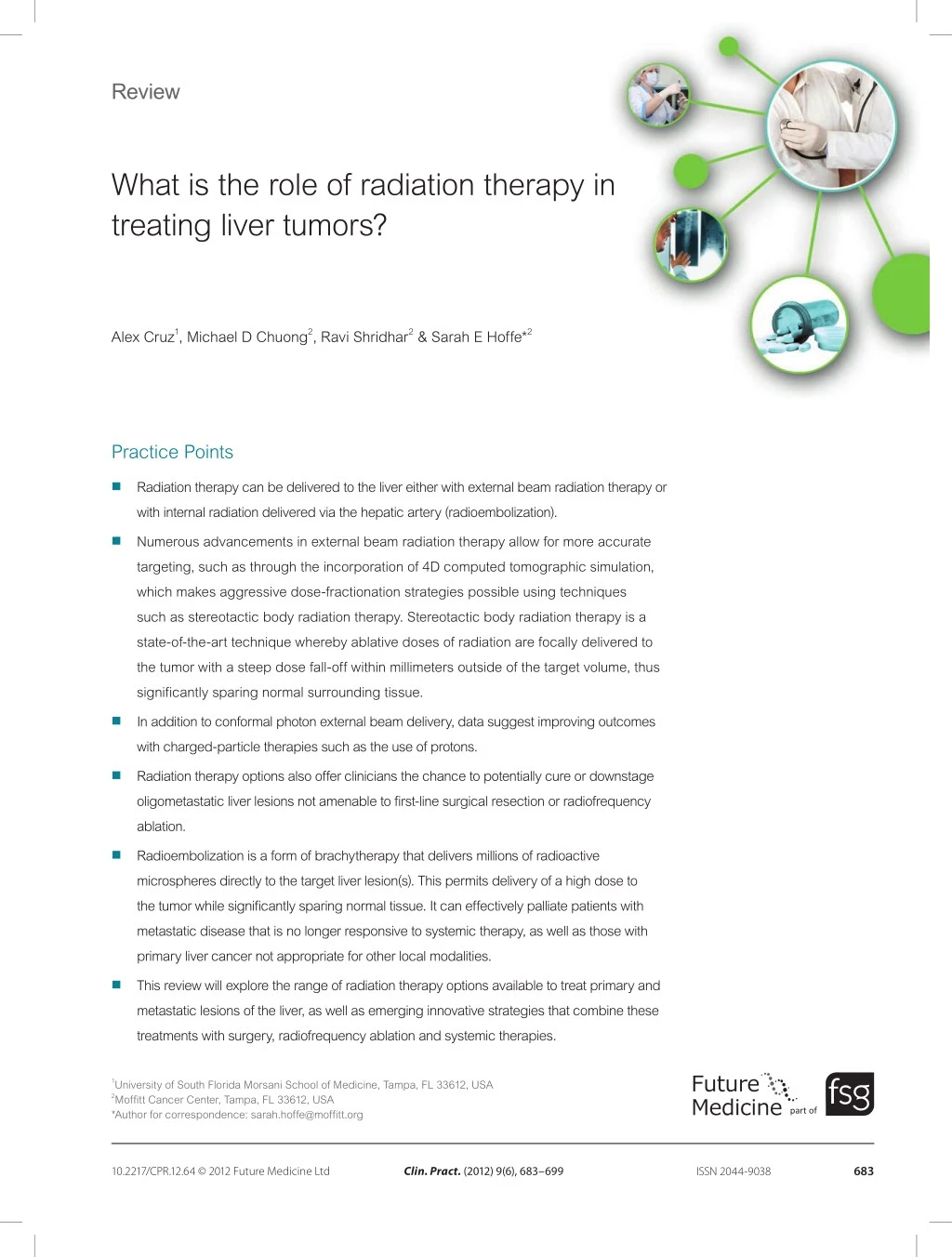
Radiation therapy can be delivered to the liver either with external beam radiation therapy or<br>with internal radiation delivered via the hepatic artery (radioembolization).

E N D
Review What is the role of radiation therapy in treating liver tumors? Alex Cruz1, Michael D Chuong2, Ravi Shridhar2 & Sarah E Hoffe*2 Practice Points Radiation therapy can be delivered to the liver either with external beam radiation therapy or with internal radiation delivered via the hepatic artery (radioembolization). Numerous advancements in external beam radiation therapy allow for more accurate targeting, such as through the incorporation of 4D computed tomographic simulation, which makes aggressive dose-fractionation strategies possible using techniques such as stereotactic body radiation therapy. Stereotactic body radiation therapy is a state-of-the-art technique whereby ablative doses of radiation are focally delivered to the tumor with a steep dose fall-off within millimeters outside of the target volume, thus significantly sparing normal surrounding tissue. In addition to conformal photon external beam delivery, data suggest improving outcomes with charged-particle therapies such as the use of protons. Radiation therapy options also offer clinicians the chance to potentially cure or downstage oligometastatic liver lesions not amenable to first-line surgical resection or radiofrequency ablation. Radioembolization is a form of brachytherapy that delivers millions of radioactive microspheres directly to the target liver lesion(s). This permits delivery of a high dose to the tumor while significantly sparing normal tissue. It can effectively palliate patients with metastatic disease that is no longer responsive to systemic therapy, as well as those with primary liver cancer not appropriate for other local modalities. This review will explore the range of radiation therapy options available to treat primary and metastatic lesions of the liver, as well as emerging innovative strategies that combine these treatments with surgery, radiofrequency ablation and systemic therapies. 1University of South Florida Morsani School of Medicine, Tampa, FL 33612, USA 2Moffitt Cancer Center, Tampa, FL 33612, USA *Author for correspondence: sarah.hoffe@moffitt.org part of 683 Clin. Pract. (2012) 9(6), 683–699 10.2217/CPR.12.64 © 2012 Future Medicine Ltd ISSN 2044-9038
Review | Cruz, Chuong, Shridhar & Hoffe summary With the liver’s dual blood supply and the preferential tumor vasculature from the hepatic artery, liver malignancies offer unique challenges and opportunities for treatment. In the case of primary liver tumors, such as hepatocellular carcinoma and cholangiocarcinoma, the disease burden, when diagnosed, is often extensive and precludes other local forms of therapy. External beam radiation therapy and intravascular brachytherapy, or radioembolization, have been shown to improve outcomes in these patients. Even though many will respond to radiation therapy, long-term cure is unlikely unless they ultimately proceed to resection or transplant. Oligometastases can also be effectively treated with radiation therapy. This review will explore the potential for radiation therapy options to be considered for both primary and metastatic liver malignancies. Liver malignancies include primary hepato cellular carcinoma (HCC), primary intrahepatic cholangiocarcinoma (IHC) and metastatic dis ease. While metastatic lesions are the most com mon form of hepatic malignancy [1,2],HCC is the sixth most common malignancy in the world and the third most common cause of cancer related mortality [3,4]. Owing to the morbidity and mortality of liver malignancies, significant effort has been invested in improving treatment techniques, especially since many patients are not optimal surgical candidates due to the extent of disease and/or poor liver function. Patients who are not optimal candidates for hepatic resection can often be selected for a pro cedure called radiofrequency abalation (RFA), which destroys tumor cells via necrosis through high temperature thermal ablation [5].Because RFA can be performed percutaneously by an interventional radiologist, or intraoperatively in collaboration with a surgeon via imageguided or manual techniques [6], a multidisciplinary tumor board will often first discuss whether either of these techniques is feasible based on the imag ing findings. Since patients often present with advanced disease with intrahepatic locations adjacent to vasculature, margin negative (R0) resections are often not feasible.Such locations are also a contraindication for RFA because it would not be possible for complete eradication due to the heatsink effect of the nearby vascula ture. To obviate the heat sink, a newer technique called irreversible electrocorporation has been developed [7]. Few data are currently available with longterm outcomes for irreversible electro corporation.There are several methods of evalu ating the effect of locoregional therapies that are outside the scope of this review [8]. These nonsurgical options are secondary to the surgical gold standard.Over the past 20 years, surgical series have demonstrated excel lent longterm survival and cure rates for patients with limited hepatic disease who undergo surgi cal management including resection or trans plant.This review will focus on the evolution and current practices of radiation therapy in treating these types of tumors in patients who are not candidates for firstline surgery or RFA. Radiation therapy can be delivered using tradi tional external beam radiation therapy (EBRT) methods or with newer techniques including stereotactic body radiation therapy (SBRT), charged particle therapy or radioembolization. The integration of new technologies has spurred renewed interest in liver tumor radiotherapy with evolving literature to support its efficacy. These advances have been critical in the pursuit of local control or potential cure. Radiation therapy modalities The use of EBRT has expanded over the course of the last several decades.Initially, EBRT was primarily used for palliation due to concern over radiationinduced wholeliver toxicity [9]. Radiationinduced liver disease (RILD), which was historically called ‘radiation hepatitis’, can potentially cause liver failure and even death. RILD classically presents with anicteric hepato megaly, elevated liver enzymes from 2 weeks to 4 months postradiation, fatigue and ascites. Occlusion and congestion of the central veins of hepatic lobules can occur while larger veins are spared [10].In addition, there is a form of non classic RILD that typically occurs from 1 week to 3 months after therapy.It is associated with EBRT 684 Clin. Pract. (2012) 9(6) future science group
What is the role of radiation therapy in treating liver tumors? | Review an elevation of liver transaminases more than fivetimes the upper limit of normal levels, or a decline in liver function, such as a decline in the Child–Pugh score by at least two [11]. One of the earliest trials to evaluate the effi cacy of radiation to the liver was performed by the Radiation Therapy Oncology Group [12]. TheRadiation Therapy Oncology Group 8003 randomized 214 patients with liver metastases to wholeliver EBRT alone (21 Gy in seven frac tions) with or without the radiosensitizer misoni dazole.While adding misonidazole did not offer a significant benefit over EBRT alone, EBRT appeared to provide a significant benefit.In fact, 77% of patients had a decrease in analgesic requirements, 67% of the patients had a decrease in abdominal distension and 40% of patients had a decrease in nausea, anorexia and vomiting. The advancement from 2D to 3D com puterized treatment planning techniques was instrumental in being able to deliver highly conformal doses to the target lesion(s) instead of uniform doses to the whole liver.Selective partial organ volumetric treatment using 3D planning software led to doseescalation stud ies.Dawson et al. published a series of over 180 patients showing that while the whole liver cannot tolerate high doses of radiation, partial liver doses can be safely escalated without induc ing RILD [13].They found that patients were at a 5% risk of RILD for uniform irradiation of onethird of the liver, twothirds of the liver and the whole liver at 90, 47 and 31 Gy, respectively [13,14].Dawson and Ten Haken also reported in a later study, the differences in wholeliver toler ance in the setting of primary versus metastatic liver cancers [14].A 5% risk of RILD occurred at a mean liver dose of 28 Gy at 2 Gy per fraction for primary liver tumors, while the same risk for RILD was seen at a mean liver dose of 32 Gy at 2 Gy per fraction for metastatic disease. Since the liver follows the parallel archi tecture model of radiobiology, it is thus desir able to treat focal regions of the liver to high tumor icidal doses, while respecting the volume of liver receiving lowdose radiation to avoid toxicity [11].Modern radiation dose limits to the liver have been founded on the principles of hepatic resection; the surgical literature notes that 75–80% of a noncirrhotic liver can be safely resected [15].However, patients with HCC are known to have impaired parenchymal func tion and liver regeneration [16].Surgeons thus perform volumetric measurements of total liver volume, as well as an estimation of the volume of the estimated future liver remnant.Similarly, radiation oncologists must determine if the vol ume of liver irradiated can be restricted such that there will be an adequate volume that is not receiving significant dose. Finally, based on Quantitative Analyses of Normal Tissue Effects in the Clinic, Pan et al. have made the following recommenda tions when treating HCC with SBRT: a mean liver dose of <13 Gy over three fractions, <18 Gy over six fractions, or <6 Gy in 4–6 Gy per fraction for classical and nonclassical RILD in Child–Pugh Class B patients[17].For patients with noncirrhotic livers treated for metastases: a mean liver dose of <15 Gy over three frac tions and <20 Gy over six fractions.In addi tion, ≥700 ml of normal liver was recommended to receive ≤15 Gy in three to five fractions [17]. SBRT Originally developed for the treatment of intra cranial malignancies, stereotactic radiotherapy has since been adopted for the treatment of extracranial disease and is called SBRT.SBRT has been most extensively evaluated for the treat ment of early stage lung cancer, but has more recently been studied for incorporation into treatment of gastrointestinal malignancies. SBRT is safely able to deliver a high ablative dose per fraction to small target volumes in only one to five fractions compared with standard fractionation treatment that delivers a much reduced dose per fraction (1.8–2 Gy) over sev eral weeks.It is thought that SBRT may poten tially have a unique radiobiologic effect com pared with standard fractionation treatment. While the exact mechanism of action is not well understood, preclinical data suggest that ablative dosing occurs at a threshold of 8–10 Gy and is due to an apoptotic effect on the vascular endothelium [18]. Accurate treatment delivery is vital for all patients, but particularly for SBRT patients because such large doses are delivered over five fractions or less. Accuracy within several milli meters can be achieved by using a combination of immobilization devices, image guidance and techniques designed to account for respiratory tumor motion, such as 4D computed tomo graphy (CT) simulation, respiratory gating, abdom inal compression and the breathhold 685 www.futuremedicine.com future science group
Review | Cruz, Chuong, Shridhar & Hoffe technique, which will be described further in the next section. Figure 1 illustrates an SBRT treat ment plan for a patient with metastatic colon cancer of the liver. can manipulate breathing patterns by restricting air entry at specified intervals [9]. Charged particle radiation therapy While the majority of EBRT is delivered using photons, other particles, such as protons or carbon ions, can be used due to their unique physical and dosimetric properties.Protons, in contrast to photons, produce no exit dose due to the Bragg–Peak effect, which can significantly limit normal tissue dose [19].Since patients with HCC typically have co existing primary liver disease, avoidance of toxicity by minimizing the volume of liver irradiated is of significant concern.Liver toxicity can also manifest itself with the reactivation of viral hepatitis in those patients with hepatitis Bassociated HCC [20]. Carbon ion particle therapy has more radio biological benefit than protons or photons, capa ble of more effectively killing hypoxic cells [21]. Both protons and carbon ions constitute a type of external beam radiation modality known as charged particles, which has a higher bio logical effectiveness than photons.These treatments hold significant promise for liver malignancies, especially HCC, given the potential for enhanc ing focaldose escalation without increasing the volumes of liver receiving lowdose radiation that could precipitate liver failure [22].Data from Asia is emerging on the incorporation of charged particle therapies for the treatment of HCC with results of 5year local control over 80% and overall survival over 35% [23].In fact, clinical results of 386 tumors treated in Japan with particle therapies in a series of 343 consecu tive patients showed a 5year local control rate of 90.8% and a survival rate of 38.2% [24]. Image-guided radiation therapy Imageguided radiation therapy can be used to assess the accuracy of the treatment setup prior to treatment delivery using either 2D (i.e., orthogo nal xrays) or 3D (i.e., conebeam CT) methods [9].Positional shifts accurate to the millimeter level can then be made to place the patient in the appropriate treatment position.4D CT simulation allows physicians to visualize tumor movement throughout the respiratory cycle with the patient being immobilized in the treatment position.Treatment volumes can then be cre ated based on the extent of tumor motion to maximize coverage of the target volume while minimizing the dose to normal surrounding structures.For patients whose tumors move a significant distance with breathing, dose to normal tissues can be minimized using several methods including abdominal compression, respiratory gating and breathhold techniques. Abdominal compression is achieved by placing a device over the abdomen to limit diaphragmatic excursion.Respiratory gating can be used in conjunction with fiducial markers during which the treatment machine will turn on within a pre determined portion of the respiratory phase, typically during end expiration.The breathhold technique monitors the patient’s breathing and Radioembolization Radioembolization is the percutaneous intra arterial injection of micronsized radio active particles that become embedded within the tumor and deliver a highfocal dose [1]. Radioembolization for primary or metastatic liver lesions is based on the dual blood supply of the liver and the preferential supply of liver lesions by the hepatic artery, whereas normal hepatocytes receive the majority of blood from the portal vein [25–28].This difference permits targeted delivery of radioactive particles to the tumor while largely sparing the normal liver [29]. Figures 2–4 highlight the benefit of radio embolization as an option for downstaging to Figure 1. Stereotactic body radiation therapy plan to a posterior liver tumor target treated with arc therapy. 686 Clin. Pract. (2012) 9(6) future science group
What is the role of radiation therapy in treating liver tumors? | Review margin negative resection; the case depictedillus trates a young patient with a solitary colo rectal metastasis who remains disease free following radioembolization and hepatic resection. Liver radioembolization is most commonly performed using yttrium90 (90Y), which is a pure bemitter, with a halflife of 64.2 h and an average energy of 0.9367 MeV.90Y decays to stable zirconium.Commercially available out patient treatment systems include the glassbased TheraSphere (Nordion, ON, Canada) and the resinbased SIRSphere (Sirtex, north Sydney, Australia).Studies have compared these two sys tems, although there is no clear consensus that one is superior to the other [30]. HCC HCC accounts for approximately 90% of all pri mary liver malignancies [9]. Hepatitis C infection is the most common etiology in North America, Japan and Europe, with 2–8% of chronically infected individuals being diagnosed with HCC each year [31]. This is in contrast to hepatitis B infection, which is the most common risk factor of HCC in Asia and Africa [32].Other risk factors include hemochromatosis, a1antitrypsin defi ciency, autoimmune hepatitis, primary biliary cirrhosis and alcoholism [31,33].Liver cirrhosis is also a wellestablished risk factor of HCC.It is estimated that upwards of 80% of HCC patients have cirrhosis as a comorbidity [34]. Since surgical resection or transplantation are potential curative options for limited HCC, the first priority is to determine the local extent of disease and classify the functional capacity of the diseased liver.Globally, there are a variety of dif ferent staging systems and liver functional classi fication systems [35]. The tumor node metastasis staging system has been adopted by the United Network for Organ Sharing (UNOS) for the staging of HCC patients [36].In this system, patients eligible for transplant are T2 (solitary tumor with vascular invasion or multiple tumors of <5 cm) or less.Moreover, the model for end stage liver disease has been adopted by UNOS to stratify patients on the liver transplant wait ing list according to the risk of death within 3 months [37]. The model for endstage liver disease score assigns points for abnormal bili rubin, creatinine and international normalized ratio values and ranges from six (less ill) to 40 (seriously ill).The Child–Pugh scoring system is another commonly used metric to evaluate Figure 2. CT scan of liver status postchemotherapy. This image shows a solitary colorectal metastasis. the patient’s clinical status [38,39].It integrates a score based on five different clinical signs (total bilirubin, prothrombin time, ascites, hepatic Figure 3. Spheres within the liver. Margin-negative resection specimen shows the embolized spheres. 687 www.futuremedicine.com future science group
Review | Cruz, Chuong, Shridhar & Hoffe While surgical resection is a firstline treat ment option for HCC, liver transplantation has gained acceptance as a definitive treatment. Longterm outcomes after liver transplantation for earlystage HCC are excellent.Mazzaferro et al. published 4year overall survival and recur rencefree survival of 85–92% in patients with either a solitary HCC of <5 cm or no more than three tumor nodules of ≤3 cm and absent vas cular or extrahepatic spread (Milan criteria) [46]. Cardenes reported a 5year survival rate of 70% for patients who underwent liver transplantation using the Milan criteria [41]. Surgical resection and/or orthotopic liver transplantation are not always the most optimal due to either unresectable disease or a shortage of donor livers.There are several nonsurgical treatment options.RFA has been used for local tumor control and as a bridge to transplanta tion [41].Other nonsurgical options include radiation therapy, transhepatic arterial chemo embolization (TACE), percutaneous ethanol injection, cryotherapy and highintensity focused ultrasound [9]. Figure 4. CT scan of liver post-treatment. No evidence of disease 1-year post-chemotherapy/spheres/surgery. EBRT EBRT has been shown to be effective and well tolerated for patients who are not optimal sur gical candidates.Liu et al. evaluated a series of 44 patients with unresectable HCC that received EBRT at a median dose of 50.4 Gy [47].The response rate was 61.4% for tumors of >5 cm. Overall survival at 12, 24 and 36 months was 60.5, 40.3 and 32.0%, respectively.The median survival was 15.2 months.BenJosef et al. found in a Phase II trial that the addition of concurrent hepatic artery floxuridine for unresectable intra hepatic malignancies with 3D conformal radia tion therapy delivered twicedaily at 1.5 Gy up to 90 Gy was associated with a 15.2month median survival for those patients with HCC with acceptable toxicity. Moreover, the study showed that total dose was the only significant predictor of survival, with little effect of dose below 60 Gy but then a steady increase in survival as the radia tion dose was escalated to 90 Gy [48].In addition, Mornex et al. noted the feasibility and efficacy of highdose 3D conformal radiation therapy in cirrhotic patients in a report of a French Phase II trial, citing a tumor response of over 90% [49].A doseescalation study from South Korea of 158 patients with primary HCC showed a dose–response relationship [50].Response rates encephalopathy and serum albumin), grading each on a scale from one to three, with one being the mildest condition and three being the most severe.Child–Pugh A, B and C classifications correspond to cumulative scores of 5–6, 7–9 or 10–15, respectively.Child–Pugh A is associated with the best prognosis while Child–Pugh C is associated with the worst. Levy and Sherman provide further information on various stag ing systems for liver malignancies [40].Table 1 compares the different staging systems and the parameters upon which they are based. Surgical resection is offered to select patients based on specific criteria.If a HCC patient pres ents with cirrhosis, they may still be a candidate for surgical resection as long as they have normal bilirubin, adequate hepatic reserve, absence of portal hypertension and disease that does not invade major vasculature, such as the inferior vena cava [41].Unfortunately, only up to 30% of patients initially present with resectable HCC and without evidence of distant metastasis [42]. Even for patients that have undergone surgical resection, the probability of local recurrence after 5 years is as high as approximately 75% [31,43], while overall survival rates range between approximately 30 and 50% [44,45]. 688 Clin. Pract. (2012) 9(6) future science group
What is the role of radiation therapy in treating liver tumors? | Review [129] [130] [131] [133] [134] [135] Ref. [132] using <40, 40–50 and >50 Gy were 29.2, 68.6 and 77.1%, respectively.Moreover, radiation dose was the only significant factor for predict ing an objective response.Data such as these have led investigators to predict that HCC tumors are indeed radiosensitive [51]. Clinically, patients with advanced HCC often present with tumor thrombus, often involving the portal vein, with reports of invasion up to 42% [52].This type of major vascular invasion is associated with a worse prognosis, with untreated patients having a median survival time of 2.7–4.0 months [43].EBRT has been reported to be effec tive in this setting [53].Investigators from Asia reported a 10.6month median survival when the radiotherapy volume included the portal vein tumor thrombus and the primary intra hepatic HCC was managed by TACE [54], and a 17.4month median survival for a thrombus location in the inferior vena cava [55]. BCLC: Barcelona Clinic Liver Cancer; CLIP: Cancer of the Liver Italian Program; CUPI: Chinese University Prognostic Index; GRETCH: Groups d’Etude et de Traitement du Carcinoma Hepatocellulaire; INR: International Normalized Performance status Symptoms a-fetoprotein thrombosis Portal vein SBRT SBRT, which delivers very high doses over one to five fractions to small volumes, has more recently been evaluated for use in HCC [17,56–59].Tse et al. published the Princess Margaret experi ence of 31 patients with Child–Pugh A HCC that were deemed unsuitable for standard thera pies [57].These patients received a median dose of 36 Gy (24–54 Gy) over six fractions.The median overall survival was 11.7 months and no patient experienced RILD.The median tumor volume was 173 ml.Data from Asia also support the safety and efficacy of SBRT [60,61]. SBRT has been evaluated as a means to bridge to transplantation. In a study reported by O’Connor et al. from the Baylor University Medical Center, ten patients were treated with SBRT to a median dose of 51 Gy in three frac tions followed by liver transplantation [58]. The median size of the 11 HCCs in this study was 3.4 cm (range = 2.5–5.5 cm) with a median followup of 62 months. The overall survival rate and diseasefree survival rate were both 100% at 5 years. Explant pathology showed a complete response rate of 27% with no viable tumor in three out of the 11 tumors. The other eight tumors were stable or had decreased. The treatment was tolerated with minimal toxicity. In a SBRT Phase I doseescalation trial, inves tigators from Indiana University (USA) reported differences in toxicity experienced by 17 patients with either Child–Pugh Class A or B [59]. Tumor extent/ stage phosphatase Alkaline Encephalopathy Table 1. Selected staging systems in hepatocellular carcinoma. Ratio; JIS: Japanese Integrated System; TNM: Tumor node metastasis treatment. Ascites INR Bilirubin Reproduced with permission from [136]. Albumin Parameter GRETCH Okuda BCLC CUPI TNM CLIP JIS 689 www.futuremedicine.com future science group
Review | Cruz, Chuong, Shridhar & Hoffe Starting at 36 Gy in three fractions, the dose was escalated in 2 Gy per fraction increments, ending at 48 Gy over three fractions.None of the patients with Child–Pugh Class A had doselimiting toxicities, while two with Child–Pugh Class B developed grade three liver toxicities when the dose was escalated to 42 Gy over three frac tions.For the entire cohort of patients, the complete response, partial response and stable disease rates were 25, 56 and 19%, respectively. At a median followup of 24 months, the local control rate was 100%.In fact, the 12month overall survival for Child–Pugh Class A was 100%, while the Child–Pugh Class B 12month overall survival was 60%.Six patients in the study were able to undergo orthotopic liver transplant ation after SBRT.Two patients had patho logical complete responses in the explanted livers, three had a partial response and one had stable disease. Cardenes et al. also reported no doselimiting toxicities in Child–Pugh Class B patients when they were treated with a dose regimen of 40 Gy over five fractions compared with 42 Gy over three fractions received by the Child–Pugh Class A patients [59]. Recently, Andolino et al. updated the Indiana University experience, reporting outcomes on 60 patients with liverconfined HCC [56].Of these patients, 36 were Child–Pugh Class A with a median number of fractions, dose per fraction and total dose of 3, 14, and 44 Gy. There were 24 patients treated with Child–Pugh Class B with a median number of fractions, dose per fraction, and total dose of 5, 8 and 40 Gy, respectively. In this series, 23 patients underwent transplantation with a median time to trans plant of 7 months.There were no grade three or greater non hematologic toxicities.The 2year local control rate was 90% with a progression free survival of 48% and an overall survival of 67%.The median tumor volume in this updated series was 27 ml. unresectable HCC [65]. The findings of minimal toxicity, an overall survival of 23.5% and a 5year local control rate of 87% are provocative.In this dataset, the subset of Child–Pugh A patients with solitary tumors had a 5year survival rate of 53.5%.Other groups have reported similar findings, suggesting that longterm survival may be possible with noninvasive therapies [63,64,66] Radioembolization Radioembolization, a form of intravascular brachytherapy, is a safe and effective treatment for HCC [51,67,68]. Like EBRT, radioembolization can also be used for tumor downstaging, stabi lization of disease as a bridge to transplantation or palliation [46,69].In a series of 150 patients, Kulik et al. report outcomes on 35 patients who were initially staged as having unresectable T3 disease [70].After radioembolization, 56% were successfully downstaged to T2 after treatment and of these, 32% were downstaged to a tumor size of ≤3 cm.Moreover, 23% of these patients were ultimately able to undergo orthotopic liver transplant.Survival rates at 1, 2 and 3 years were 84, 54 and 27%, respectively.Data confirming the efficacy of radioembolization from a histo pathologic standpoint have been accumulating. In a study of 35 patients with 38 lesions that were treated with 90Y radioembolization prior to transplant, Riaz et al. showed that all target lesions showed some degree of histologic necro sis at explant [71]. In patients with lesions of <3 cm, 89% had complete histologic necrosis. Overall, 61% of target lesions showed complete pathologic necrosis. Prior to the widespread use of radio embolization, TACE was the predominant option for liverdirected therapy [72].Although no prospective data exist, Salem et al. conducted a comparative effectiveness study retrospectively between chemoembolization and 90Y radio embolization in a cohort of 245 patients treated at Northwestern University (IL, USA) [73]. Radioembolization was delivered to 123 patients while 122 were treated with TACE; none of the patients in either cohort had evidence of extra hepatic metastases or portal vein thrombosis. Patients receiving radioembolization had a higher response rate (49 vs 36%).The median survival for patients receiving radioembolization was also longer, although this difference was not statisti cally significant (20.5 vs 17.4 months; p = 0.232) [73,74].Salem et al. recently presented their Charged particle therapy Carbon ion therapy is emerging as a potential option in Asia.Kato et al. reported results from a Phase I/II trial that treated patients with HCC to a dose of 50–80 Gy with 5year local con trol of 81% and survival of 25% [62].Proton beam data has suggested efficacy in this setting as well [63,64].Chiba et al. have reported data from Japan with protonbeam therapy to a dose of 72 Gy in 16 fractions for 162 patients with 690 Clin. Pract. (2012) 9(6) future science group
What is the role of radiation therapy in treating liver tumors? | Review SBRT SBRT has been evaluated in the setting of IHC.Barney et al. explored the use of SBRT at Mayo Clinic (MN, USA) in a cohort of ten patients with 12 unresectable primary tumors (n = 6) or recurrent IHC (n = 6) [81].SBRT was delivered over three (n = 2) or five (n = 10) consecutive fractions to a median dose of 55 Gy (range 45–60 Gy).After a median followup of 14 months, freedom from progression within the SBRT treatment field was 100%, but four patients developed intrahepatic recurrence in other areas of the liver.Overall survival was 83% at 6 months and 73% at 12 months.Five patients experienced grade two nausea and vom iting, while one patient experienced grade five liver failure.As noted above, Tse et al. reported the Princess Margaret Hospital experience with SBRT for primary liver malignancies [57].In their series, the ten patients with IHC had a median survival of 15 months.Ibarra et al. reported a 75% 6month overall survival and a 45% 1year overall survival in a multicenter study of SBRT for nonresectable primary liver tumors with low rates of toxicity [82]. outcomes of 526 radioembolization treatments noting response rates of 42% using the WHO criteria and 57% using European Association for the Study of the Liver (EASL) criteria [75]. Patients with Child–Pugh A disease fared the best, with a median survival of 17.2 compared with 7.7 months in those with Child–Pugh B disease.Patients with Child–Pugh A disease and portal vein thrombosis benefited from treatment, but those with Child–Pugh B disease and portal vein thrombosis had a median survival of only 5.6 months.Raoul et al. compared chemoembo lization and radioembolization in a prospective randomized trial of 142 patients [74]; however, instead of 90Ybased, 131Ilabeled iodized oil (Guerbet LLC, IN, USA) was used. Overall sur vival was similar between the treatment modali ties.Overall survival rates were reported at 6, 12, 24, 36 and 48 months.For the 131Ilipiodol patients, the overall survival rates were 69, 38, 22, 14 and 10%, respectively.For the chemo embolization patients, the overall survival rates were 66, 42, 22, 3 and 0%, respectively.The number of patients achieving a complete versus partial response was similar between the groups. Cholangiocarcinoma IHC comprises approximately 10% of all primary liver malignancies.The only known curative treatment for IHC is surgical resection, which is not commonly feasible due to its aggressive and infiltrative nature [76,77].Even for those that are fortunate enough to receive surgery, local recur rence at the site of resection is common [76,78]. The prognosis for un resectable IHC, even with nonsurgical treatment, is poor, with an expected median survival rate of approximately 9 months and a 5year overall survival rate of <5% [79]. Radioembolization Radioembolization may also be a therapeutic option for patients with IHC.Ibrahim et al. reported results from Northwestern University showing a median overall survival of 14.9 months in the 24patient study [83].The median survival was improved (31.8 months) for patients who had an excellent performance status (Eastern Cooperative Oncology Group 0).Over 50% of tumor necrosis was demonstrated using the EASL criteria in 77% of patients.German inves tigators also suggest potential benefit, reporting the results in 33 patients with 90Y [84].Median overall survival was 22 months with time to progression of 9.8 months.Similar to the US study, the median survival in patients with Eastern Cooperative Oncology Group 0 was 29.4 months [84]. EBRT EBRT can be used to improve prognosis and lessen morbidity in patients with IHC. Chen et al. reported the effectiveness of EBRT in 84 patients diagnosed with unresectable IHC [80]. Of these, 35 patients received EBRT to a median dose of 50 Gy (range 30–60 Gy) in 2 Gy fractions.The median overall survival for the EBRT and nonEBRT groups were 9.5 and 5.1 months, respectively.The 1 and 2year over all survival rates for the EBRT cohort was 38.5 and 16.4%, respectively, while it was 9.6 and 4.9%, respectively, for the patients who did not receive EBRT. Metastases Treatment of liveronly metastases is of sig nificant interest due to the expanding range of treatment options.The term oligometastasis is relevant because there may be subsets of patients who only have a limited number of metastatic tumor clones that can potentially be completely eradicated.Due to the vascular nature of the 691 www.futuremedicine.com future science group
Review | Cruz, Chuong, Shridhar & Hoffe liver, it is a common site of metastasis, partic ularly from primary gastrointestinal cancers, especially for colorectal carcinomas (CRCs) [85].Approximately 25% of CRC patients have hepatic metastases at the time of diagnosis, while nearly half of all patients with CRC have tumor recurrences in the liver in the span of 5 years [86]. It is with this rationale that oncologists have found that treating liver oligometastases is poten tially curative.In the case of CRC, for example, surgical resection is associated with the potential for a longterm cure.The 5year overall survival rates for selected patients with resected CRC liver metastases are approximately 60% [87–93]. A 10year survival rate has also indicated that surgical resection is potentially curative, with multiple institutions reporting overall survival of up to approximately 30% [88,91,92,94–97].In terms of surgical resection for CRC liver metas tases, Fong et al. have reported that patients with a solitary tumor of <5 cm, long diseasefree interval (>1 year), carcinoembryonic antigen of <200 ng/ml and negative surgical margins had 5year overall survival of 60% [88].Patients who did not meet these criteria had 5year OS of only 14% [88].In this series, patients were not treated with prior systemic chemotherapy.While a large body of literature has been collected on CRC oligo metastases over the past 30 years, there are also documented cases of longterm survival from liver oligometastases originating from nonCRCs, such as breast cancers and sar comas, although treatment options for nonCRC metastases are less defined [98]. As 80–90% of patients with metastatic liver disease are considered unresectable, due to either large tumor size or location, nonsurgical inter ventions are the only options available for these patients [99].The possibility of curative treatment for liver metastases raises the question of whether other treatment options can provide similar results to surgical resection when resection is not possible.If so, then what is the maximum number and size of tumors before the chance of survival decreases? Also, what role does tumor location provide in noninvasive therapies? In patients with liver metastases not appro priate for surgery, RFA is often considered by multidisciplinary tumor boards as a potential option.Aloia et al. performed an analysis com paring the efficacy of RFA against surgical resec tion in the setting of solitary CRC liver metastasis [100].They noted that RFA, while having similar rates of intrahepatic and extrahepatic failure, had a seventimes higher risk of local failure. The risk of death was also threetimes higher with RFA [100].A study by Otto et al. reported a local failure rate of 32%, with a 12month local control rate of 58% and a 12month rate of hepatic retreatment of 33% in 28 patients with CRC liver oligo metastases [101]. Finally, a study conducted by Livraghi and Solbiati found local failures occurring in 70 out of 179 patients (39%) treated with RFA for CRC liver oligometastases, with an 18month local control rate of 56% [102]. There has also been a trend of decreasing macro scopic local recurrence with smaller tumor size in patients treated with percutaneous RFA. In 2001, Solbiati et al. reported that, in a series of 117 patients with 179 tumors with an average size of 2.8 cm, the patients had a macroscopic local recurrence rate of 39% [103].In 2006, Solbiati et al. reported that, in a series of 121 patients with 320 tumors with an average size of 2.1 cm, the macroscopic local recurrence rate was 14% [104]. Although RFA can effectively provide improved local control in liver oligo metastases, RFA may not be optimal due to tumor size and location. EBRT Safety and efficacy data supporting the role of conformal partial liver EBRT for the treatment of metastatic colorectal carcinomas was reported by investigators at the University of Michigan (USA) in the 1990s [105].Robertson et al. noted a response rate of 50% in patients treated with hyperfractionated 3D conformal radiation therapy to a maximum dose of 72.6 Gy in frac tions of 1.50–1.65 Gy [105].In this 22patient study, the median survival was 20 months.The Michigan group has also reported that higher doses are associated with improved median survival in patients with colorectal cancer metastatic to the liver [48,106]. They noted a 17.2month median survival rate in a series of 47 patients who were treated at 1.5 Gy twice daily to a median dose of 60.75 Gy along with concurrent continuousinfusion hepaticarterial floxuridine [48]. SBRT The SBRT literature for the treatment of liver metastases has been expanding with an evolv ing foundation of clinical outcomes data [107]. One of the first SBRT studies for liver metastases was conducted at the University of Heidelberg 692 Clin. Pract. (2012) 9(6) future science group
What is the role of radiation therapy in treating liver tumors? | Review (Germany), where patients were treated with 14–26 Gy in a single fraction.Forty three of the 55 lesions treated were locally controlled at a median followup of 5.7 months. Furthermore, by using a single fraction of 22 Gy, Herfarth et al. were able to show a local control rate of 66% after 18 months [108,109].Hoyer et al. also concluded that SBRT is reasonable for treatment with curative intent in a series of 64 patients with 141 CRC tumors (44 of which were un resectable) [110]. While SBRT can be used for palliation, it may also be used to bridge poten tially curable patients with oligometastases to complete disease eradication.Adam et al. have shown that downstaging of CRC liver metastases to allow tumor resection has a survival benefit, especially in a population of patients that have been pretreated with chemotherapy [111]. A multiinstitutional, prospective study con ducted by Chang et al. at Princess Margaret Hospital (ON, Canada), University of Colorado (USA) and Stanford University (CA, USA) attempted to establish the standard of care ther apy for patients with CRC metastases of the liver using SBRT by determining outcomes of a large patient cohort [112].In their study, they found that active extrahepatic disease correlated with a decrease in overall survival time (p = 0.04), coinciding with surgical studies that have established active extrahepatic disease correlat ing with worse survival in patients under going hepatic resection [113–116].This study limited the maximum number of tumor lesions to four, while having a solitary lesion versus two to four lesions did not provide any survival benefit [112]. They formulated that a threefraction regimen to a total dose of 48 Gy provided optimal ablative therapy while minimizing radiation toxicity to the liver and surrounding normal tissue. Recently, Rusthoven et al. have reported their experiences from a multiinstitutional Phase I/II trial with 47 patients with 63 lesions treated with SBRT [117].The patients contained one to three lesions, with 6 cm as the individual maximal tumor size allowed.By delivering a total dose of 60 Gy over three fractions, they were able to maintain a 100% local control rate in the patients with tumor sizes of ≤3 cm.However, one patient in the study suffered from grade three or higher toxicity.The median individual maximal tumor diameter was 2.7 cm and the median overall survival was 20.5 months, while the 24month overall survival rate was 30%. The main concern with SBRT to the liver is hepatotoxicity. Radiation oncologists are con servative in their dose–volume constraints when treating liver tumors because of concerns of caus ing RILD. To be safe, a standard guideline of 700 ml of normal liver tissue receiving no more than 15 Gy delivered over three fractions (or 7 Gy in one fraction) has been established in order to minimize toxicities [118]. Also, radiation oncologists must try to keep the radiation dose low enough, such that 30% of the liver volume receives no more than 21 Gy in three fractions (or 12 Gy in one fraction) [119,120]. Radioembolization Radioembolization for liver metastases is another viable alternative to surgical resection [121].Kennedy et al. reported a modern US expe rience with a cohort of 208 heavily pretreated patients with metastatic colorectal liver metas tases, reporting an encouraging median survival of 10.5 months for responders [122].In a series of patients treated at Northwestern University and William Beaumont Hospital (MI, USA), Sato et al. reported on a cohort of 51 patients with CRC liver metastases who received 90Y radioembolization [123]. Median survival time was 15.2 months.Mulcahy et al. reported that 90Y radioembolization for liver metastases resulted in median survival of 14.5 months from time of initial treatment and a time to hepatic progression of 15.4 months after initial treat ment [124].More recently, Nace et al. reported a median survival of 17 months in patients without extrahepatic disease and an even lon ger median overall survival of 18.3 months in the patients without extrahepatic disease that had not received cetuximab prior to radio embolization [125].The patients in this series had metastatic CRC and were not candidates for surgical resection or RFA, and had received either first or secondline chemotherapy. There has been interest in exploring com binations of systemic chemotherapy with radio embolization.Two randomized con trolled Phase III trials showed benefits for SIR SPHERES combined with chemo therapy com pared with chemotherapy alone in patients with CRC liver metastases [126,127].With more effec tive systemic therapies that have been introduced into the metastatic CRC paradigm, future stud ies will continue to evaluate potential efficacy of combined modality regimens. 693 www.futuremedicine.com future science group
Review | Cruz, Chuong, Shridhar & Hoffe Discussion Over the last 20 years, significant progress in the localization of hepatic tumors that move with respiration and the careful delivery of focal highdose EBRT have created new opportunities for noninvasive liver treatment.Technological advances are now leading the way for improved outcomes, with studies documenting doserelated efficacy with low toxicity.Internal radiation with intravascular microsphere brachy therapy has also emerged as a welltolerated, effective treatment, with a potential role in downstaging to surgery or RFA, as well as palliating patients with large disease burden.Table 2 summarizes key studies and survival outcomes for patients with primary or metastatic liver disease using different radiotherapy techniques. optimal number and size of lesions to be con sidered for a potentially curative, tumoricidal approach? The case with primary liver malignancies is especially unclear.Focal irradiation of HCC lesions is associated with high infield control rates but progression outside of the treatment field is common [128].How best to optimize liver directed therapies potentially capable of treating microscopic disease in the whole lobe, along with focal EBRT to the macroscopic disease, is an important question.Given the promising chargedparticle data, what is the role of radiation sensitizers to improve the excellent 5year out comes even further?Is a longterm cure possible without surgery, transplantation or RFA? Finally, the future possibilities of how best to integrate radioembolization into the exist ing treatment paradigm for liver malignancies is expanding.With the potential of delivering selective treatment based on tumor blood sup ply, radiation oncologists have the ability to deliver extremely high doses to a small volume, such as a particular liver segment.Ablation of a focal liver segment by radioembolization could potentially become a standalone, curative treat ment.This could yield more options for patients with isolated disease that may not be feasible for resection or RFA due to tumor location within the liver.As systemic therapies continue to evolve and improve, future trials are needed to explore novel combinations of local radiation treatment with such agents. Conclusion & future perspective Patients with primary and metastatic hepatic tumors can significantly benefit from liver directed radiation therapy.Despite the recent advances in radiation treatment delivery, there are still many unanswered questions that will hopefully be answered over the next 5–10 years. For metastatic lesions of the liver, the ques tion of a potential cure for oligometastases is indeed appealing.Optimal patient selection, however, is unclear.When should patients who have limited hepatic metastases with a favorable primary cancer be considered for external radia tion modalities?How should systemic therapy be integrated with such treatment?What is the Table 2. Summary of survival outcomes for patients with primary or metastatic liver cancer who received radiation therapy. Study (year) Salem et al. (2007) Liu et al. (2004) Ben-Josef et al. (2005) Tse et al. (2008) Cardenes et al. (2010) Chiba et al. (2005) Chen et al. (2010) Barney et al. (2012) Ibrahim et al. (2008) Hoffmann et al. (2012) Sato et al. (2008) Nace et al. (2011) Van Hazel et al. (2004) Rusthoven et al. (2009) Chang et al. (2011) CRT: Conformal radiation therapy; HCC: Hepatocellular carcinoma; IHC: Intrahepatic cholangiocarcinoma; Met: Metastasis; NR: Not reported; SBRT: Stereotactic body radiation therapy. Patients (n) 123 44 35 31 17 162 35 10 24 33 51 51 11 38 47 Histology HCC HCC HCC HCC HCC HCC IHC IHC IHC IHC Met Met Met Met Met Concurrent chemotherapy No No Yes No No No No No No No No No Yes No No Technique Radioembolization 3D-CRT 3D-CRT SBRT SBRT Proton therapy 3D-CRT SBRT Radioembolization Radioembolization Radioembolization Radioembolization Radioembolization SBRT SBRT Median survival (months) 20.5 15.2 15.2 11.7 NR 26.4 9.5 NR 14.9 22 15.2 18.3 29.4 20.5 14.4 Ref. [28] [47] [48] [57] [59] [65] [80] [81] [83] [84] [123] [125] [127] [117] [112] 694 Clin. Pract. (2012) 9(6) future science group
What is the role of radiation therapy in treating liver tumors? | Review Financial & competing interests disclosure The authors have no relevant affiliations or financial involve- ment with any organization or entity with a financial interest in or financial conflict with the subject matter or materials discussed in the manuscript. This includes employment, con- sultancies, honoraria, stock ownership or options, expert t estimony, grants or patents received or pending, or royalties. No writing assistance was utilized in the production of this manuscript. Since so many patients experience liver involvement as a component of their disease, the enlarging range of safe and effective radia tion options is indeed appealing.Bolstered by the evolving efficacy data of both internal and external radiation modalities, the promise of bet ter outcomes shines brighter than ever before due to more effective tools in our treatment armamentarium. Hoffe SE, Finkelstein SE, Russell MS, Shridhar R. Nonsurgical options for hepatocellular carcinoma: evolving role of external beam radiotherapy. Cancer Control 17(2), 100–110 (2010). biology? Int. J. Radiat. Oncol. Biol. Phys. 71(2), 324–325 (2008). 9 References Papers of special note have been highlighted as: of interest 19 Skinner HD, Hong TS, Krishnan S. Chargedparticle therapy for hepatocellular carcinoma. Semin. Radiat. Oncol. 21(4), 278–286 (2011). Memon K, Lewandowski RJ, Kulik L, Riaz A, Mulcahy MF, Salem R. Radioembolization for primary and metastatic liver cancer. Semin. Radiat. Oncol. 21(4), 294–302 (2011). 1 10 Dawson LA, Normolle D, Balter JM, McGinn CJ, Lawrence TS, Ten Haken RK. Analysis of radiationinduced liver disease using the Lyman NTCP model. Int. J. Radiat. Oncol. Biol. Phys. 53(4), 810–821 (2002). Provides a comprehensive review of the rationale for incorporating charged particles into hepato cellular carcinoma treatment. Moreover, the data supporting the durable local control rates >80% and 5-year survival rates of 25% are reviewed. Provides the reader with the principles of radioembolization. Lewandowski RJ, Thurston KG, Goin JE et al.90Y microsphere (TheraSphere) treatment for unresectable colorectal cancer metastases of the liver: response to treatment at targeted doses of 135–150 Gy as measured by 18Ffluorodeoxyglucose positron emission tomography and computed tomographic imaging. J. Vasc. Interv. Radiol. 16(12), 1641–1651 (2005). 2 Pan CC, Kavanagh BD, Dawson LA et al. Radiationassociated liver injury. Int. J. Radiat. Oncol. Biol. Phys. 76(Suppl. 3), S94–S100 (2010). 11 20 Kim JH, Park JW, Kim TH, Koh DW, Lee WJ, Kim CM. Hepatitis B virus reactivation after threedimensional conformal radiotherapy in patients with hepatitis B virusrelated hepatocellular carcinoma. Int. J. Radiat. Oncol. Biol. Phys. 69(3), 813–819 (2007). Report of the QUANTEC group that is wide in scope and thoroughly reviews the pathophysiology of liver injury. Guidelines are provided for avoiding liver toxicity. Parkin DM, Bray F, Ferlay J, Pisani P. Global cancer statistics, 2002. CA Cancer J. Clin. 55(2), 74–108 (2005). 3 12 Leibel S, Bauer M, Wasserman T et al. Radiotherapy with or without misonidazole for patients with stage IIIB or stage IVA squamous cell carcinoma of the uterine cervix: preliminary report of a Radiation Therapy Oncology Group randomized trial. Int. J. Radiat. Oncol. Biol. Phys. 13(4), 541–549 (1987). 21 Jiang GL. Particle therapy for cancers: a new weapon in radiation therapy. Front. Med. 6(2), 165–172 (2012). Bosch FX, Ribes J, Diaz M, Cleries R. Primary liver cancer: worldwide incidence and trends. Gastroenterology 127(5 Suppl. 1), S5–S16 (2004). 4 22 Skinner HD, Hong TS, Krishnan S. Charged particle therapy for hepatocellular carcinoma. Semin. Radiat. Oncol. 21(4), 278–286 (2011). 23 Dawson LA. Protons or photons for hepatocellular carcinoma? Let’s move forward together. Int. J. Radiat. Oncol. Biol. Phys. 74(3), 661–663 (2009). Timmerman RD, Bizekis CS, Pass HI et al. Local surgical, ablative, and radiation treatment of metastases. CA Cancer J. Clin. 59(3), 145–170 (2009). 5 13 Dawson LA, Ten Haken RK, Lawrence TS. Partial irradiation of the liver. Semin. Radiat. Oncol. 11(3), 240–246 (2001). 24 Komatsu S, Fukumoto T, Demizu Y et al. Clinical results and risk factors of proton and carbon ion therapy for hepatocellular carcinoma. Cancer 117(21), 4890–4904 (2011). Thorough review of oligometastases and local treatment modalities. Emphasis on rationale and technique is helpful to the reader to provide a broad context for the emerging role of more aggressive local therapies. 14 Dawson LA, Ten Haken RK. Partial volume tolerance of the liver to radiation. Semin. Radiat. Oncol. 15(4), 279–283 (2005). Penna C, Nordlinger B. Colorectal metastasis (liver and lung). Surg. Clin. North Am. 82(5), 1075–1090, xxi (2002). 15 25 Breedis C, Young G. The blood supply of neoplasms in the liver. Am. J. Pathol. 30(5), 969–977 (1954). Gillams AR. The use of radiofrequency in cancer. Br. J. Cancer 92, 1825–1829 (2005). 16 Vauthey JN, Chaoui A, Do KA et al. Standardized measurement of the future liver remnant prior to extended liver resection: methodology and clinical associations. Surgery 127(5), 512–519 (2000). 6 26 Liu DM, Salem R, Bui JT et al. Angiographic considerations in patients undergoing liverdirected therapy. J. Vasc. Interv. Radiol. 16(7), 911–935 (2005). Narayanan G. Irreversible electroporation for treatment of liver cancer. Gastroenterol. Hepatol. 7(5), 313–316 (2011). 7 17 Pan CC, Kavanagh BD, Dawson LA et al. Radiationassociated liver injury. Int. J. Radiat. Oncol. Biol. Phys. 76(Suppl. 3), S94–S100 (2010). Riaz A, Memon K, Miller FH et al. Role of the EASL, RECIST, and WHO response guidelines alone or in combination for hepatocellular carcinoma: radiologic–pathologic correlation. J. Hepatol. 54(4), 695–704 (2011). 8 27 Lewandowski RJ, Sato KT, Atassi B et al. Radioembolization with 90Y microspheres: angiographic and technical considerations. Cardiovasc. Intervent. Radiol. 30(4), 571–592 (2007). 18 Brown JM, Koong AC. Highdose single fraction radiotherapy: exploiting a new 695 www.futuremedicine.com future science group
Review | Cruz, Chuong, Shridhar & Hoffe 28 Salem R, Lewandowski RJ, Sato KT et al. Technical aspects of radioembolization with 90Y microspheres. Tech. Vasc. Interv. Radiol. 10(1), 12–29 (2007). 42 Lau WY, Lai EC. Salvage surgery following downstaging of unresectable hepatocellular carcinoma – a strategy to increase resectability. Ann. Surg. Oncol. 14(12), 3301–3309 (2007). and computer tomography characteristics. Acta Med. Okayama 66(2), 131–141 (2012). 53 Hawkins MA, Dawson LA. Radiation therapy for hepatocellular carcinoma: from palliation to cure. Cancer 106(8), 1653–1663 (2006). 29 Riaz A, Gates VL, Atassi B et al. Radiation segmentectomy: a novel approach to increase safety and efficacy of radioembolization. Int. J. Radiat. Oncol. Biol. Phys. 79(1), 163–171 (2011). 43 Llovet JM, Bustamante J, Castells A et al. Natural history of untreated nonsurgical hepatocellular carcinoma: rationale for the design and evaluation of therapeutic trials. Hepatology 29(1), 62–67 (1999). 54 Yoon SM, Lim YS, Won HJ et al. Radiotherapy plus transarterial chemoembolization for hepatocellular carcinoma invading the portal vein: longterm patient outcomes. Int. J. Radiat. Oncol. Biol. Phys. 82(5), 2004–2011 (2012). 30 Vente MA, Wondergem M, van der Tweel I et al. Yttrium90 microsphere radioembolization for the treatment of liver malignancies: a structured metaanalysis. Eur. Radiol. 19(4), 951–959 (2009). 44 Chok KS, Ng KK, Poon RT, Lo CM, Fan ST. Impact of postoperative complications on longterm outcome of curative resection for hepatocellular carcinoma. Br. J. Surg. 96(1), 81–87 (2009). 55 Hou JZ, Zeng ZC, Zhang JY, Fan J, Zhou J, Zeng MS. Influence of tumor thrombus location on the outcome of externalbeam radiation therapy in advanced hepatocellular carcinoma with macrovascular invasion. Int. J. Radiat. Oncol. Biol. Phys. 84(2), 362–368 (2012). 31 Bruix J, Sherman M. Management of hepatocellular carcinoma. Hepatology 42(5), 1208–1236 (2005). 45 Kianmanesh R, Regimbeau JM, Belghiti J. Selective approach to major hepatic resection for hepatocellular carcinoma in chronic liver disease. Surg. Oncol. Clin. N. Am. 12(1), 51–63 (2003). 32 Bosch FX, Ribes J, Borras J. Epidemiology of primary liver cancer. Semin. Liver Dis. 19(3), 271–285 (1999). 56 Andolino DL, Johnson CS, Maluccio M et al. Stereotactic body radiotherapy for primary hepatocellular carcinoma. Int. J. Radiat. Oncol. Biol. Phys. 81(4), e447–e453 (2011). 46 Mazzaferro V, Regalia E, Doci R et al. Liver transplantation for the treatment of small hepatocellular carcinomas in patients with cirrhosis. N. Engl. J. Med. 334(11), 693–699 (1996). 33 Fattovich G, Stroffolini T, Zagni I, Donato F. Hepatocellular carcinoma in cirrhosis: incidence and risk factors. Gastroenterology 127(5 Suppl. 1), S35–S50 (2004). 57 Tse RV, Hawkins M, Lockwood G et al. Phase I study of individualized stereotactic body radiotherapy for hepatocellular carcinoma and intrahepatic cholangiocarcinoma. J. Clin. Oncol. 26(4), 657–664 (2008). 34 Bartlett DL, Dibisceglie AM, Dawson LA. Cancer of the Liver. Lippincott Williams & Wilkins, PA, USA, 1129–1156 (2008). 47 Liu MT, Li SH, Chu TC et al. Three dimensional conformal radiation therapy for unresectable hepatocellular carcinoma patients who had failed with or were unsuited for transcatheter arterial chemoembolization. Jpn J. Clin. Oncol. 34(9), 532–539 (2004). 58 O’Connor JK, Trotter J, Davis GL et al. Longterm outcomes of stereotactic body radiation therapy in the treatment of hepatocellular cancer as a bridge to transplantation. Liver Transpl. 18(8), 949–954 (2012). 35 Waly Raphael S, Yangde Z, Yuxiang C. Hepatocellular carcinoma: focus on different aspects of management. ISRN Oncol. 2012, 421673 (2012). 48 BenJosef E, Normolle D, Ensminger WD et al. Phase II trial of highdose conformal radiation therapy with concurrent hepatic artery floxuridine for unresectable intrahepatic malignancies. J. Clin. Oncol. 23(34), 8739–8747 (2005). 36 Yao FY, Bass NM, Ascher NL, Roberts JP. Liver transplantation for hepatocellular carcinoma: lessons from the first year under the Model of EndStage Liver Disease (MELD) organ allocation policy. Liver Transpl. 10(5), 621–630 (2004). 59 Cardenes HR, Price TR, Perkins SM et al. Phase I feasibility trial of stereotactic body radiation therapy for primary hepatocellular carcinoma. Clin. Transl Oncol. 12(3), 218–225 (2010). 49 Mornex F, Girard N, Beziat C et al. Feasibility and efficacy of highdose threedimensionalconformal radiotherapy in cirrhotic patients with smallsize hepatocellular carcinoma noneligible for curative therapies – mature results of the French Phase II RTF1 trial. Int. J. Radiat. Oncol. Biol. Phys. 66(4), 1152–1158 (2006). 60 Kang JK, Kim MS, Cho CK et al. Stereotactic body radiation therapy for inoperable hepatocellular carcinoma as a local salvage treatment after incomplete transarterial chemoembolization. Cancer doi:10.1002/ cncr.27533 (2012) (Epub ahead of print). 37 Martin AP, Bartels M, Hauss J, Fangmann J. Overview of the MELD score and the UNOS adult liver allocation system. Transplant. Proc. 39(10), 3169–3174 (2007). 38 Child CG, Turcotte JG. Surgery and portal hypertension. Major Probl. Clin. Surg. 1, 1–85 (1964). 61 Seo YS, Kim MS, Yoo SY et al. Preliminary result of stereotactic body radiotherapy as a local salvage treatment for inoperable hepatocellular carcinoma. J. Surg. Oncol. 102(3), 209–214 (2010). 39 Pugh RN, MurrayLyon IM, Dawson JL, Pietroni MC, Williams R. Transection of the oesophagus for bleeding oesophageal varices. Br. J. Surg. 60(8), 646–649 (1973). 50 Park HC, Seong J, Han KH, Chon CY, Moon YM, Suh CO. Dose–response relationship in local radiotherapy for hepatocellular carcinoma. Int. J. Radiat. Oncol. Biol. Phys. 54(1), 150–155 (2002). 62 Kato H, Tsujii H, Miyamoto T et al. Results of the first prospective study of carbon ion radiotherapy for hepatocellular carcinoma with liver cirrhosis. Int. J. Radiat. Oncol. Biol. Phys. 59(5), 1468–1476 (2004). 40 Levy I, Sherman M. Staging of hepatocellular carcinoma: assessment of the CLIP, Okuda, and Child–Pugh staging systems in a cohort of 257 patients in Toronto. Gut 50(6), 881–885 (2002). 51 Schwarz RE, AbouAlfa GK, Geschwind JF, Krishnan S, Salem R, Venook AP. Nonoperative therapies for combined modality treatment of hepatocellular cancer: expert consensus statement. HPB (Oxford) 12(5), 313–320 (2010). 63 Bush DA, Hillebrand DJ, Slater JM, Slater JD. Highdose proton beam radiotherapy of hepatocellular carcinoma: preliminary results of a Phase II trial. Gastroenterology 127(5 Suppl. 1), S189–S193 (2004). 41 Cardenes HR. Role of stereotactic body radiotherapy in the management of primary hepatocellular carcinoma. Rationale, technique and results. Clin. Transl Oncol. 11(5), 276–283 (2009). 52 Jia L, Kiryu S, Watadani T et al. Prognosis of hepatocellular carcinoma with portal vein tumor thrombus: assessment based on clinical 696 Clin. Pract. (2012) 9(6) future science group
What is the role of radiation therapy in treating liver tumors? | Review unresectable intrahepatic cholangiocarcinoma: factors associated with prolonged survival. Cardiovasc. Intervent. Radiol. 35(1), 105–116 (2012). 64 Kawashima M, Furuse J, Nishio T et al. Phase II study of radiotherapy employing proton beam for hepatocellular carcinoma. J. Clin. Oncol. 23(9), 1839–1846 (2005). injection of 131Ilabelediodized oil in the treatment of hepatocellular carcinoma. Hepatology 26(5), 1156–1161 (1997). 75 Salem R, Lewandowski RJ, Mulcahy MF et al. Radioembolization for hepatocellular carcinoma using Yttrium90 microspheres: a comprehensive report of longterm outcomes. Gastroenterology 138(1), 52–64 (2010). 65 Chiba T, Tokuuye K, Matsuzaki Y et al. Proton beam therapy for hepatocellular carcinoma: a retrospective review of 162 patients. Clin. Cancer Res. 11(10), 3799–3805 (2005). 85 Wang J, Yang M, Hoffman RM. Visualizing portal vein metastatic trafficking to the liver with green fluorescent proteinexpressing tumor cells. Anticancer Res. 24(6), 3699–3702 (2004). 66 Fukumitsu N, Sugahara S, Nakayama H et al. A prospective study of hypofractionated proton beam therapy for patients with hepatocellular carcinoma. Int. J. Radiat. Oncol. Biol. Phys. 74(3), 831–836 (2009). 86 Bengmark S, Hafstrom L. The natural history of primary and secondary malignant tumors of the liver. I. The prognosis for patients with hepatic metastases from colonic and rectal carcinoma by laparotomy. Cancer 23(1), 198–202 (1969). This report from the Northwestern University (IL, USA) group details the results of over 500 radioembolization treatments. The study is comprehensive in reporting toxicity, outcomes and response data. 67 Collettini F, Schnapauff D, Poellinger A et al. Hepatocellular carcinoma: computed tomographyguided highdoserate brachytherapy (CTHDRBT) ablation of large (5–7 cm) and very large (>7 cm) tumours. Eur. Radiol. 22(5), 1101–1109 (2012). 87 Simmonds PC, Primrose JN, Colquitt JL, Garden OJ, Poston GJ, Rees M. Surgical resection of hepatic metastases from colorectal cancer: a systematic review of published studies. Br. J. Cancer 94(7), 982–999 (2006). 76 Jarnagin WR, Fong Y, Dematteo RP et al. Staging, resectability, and outcome in 225 patients with hilar cholangiocarcinoma. Ann. Surg. 234(4), 507–517; discussion 517–519 (2001). 77 Rea DJ, Heimbach JK, Rosen CB et al. Liver transplantation with neoadjuvant chemoradiation is more effective than resection for hilar cholangiocarcinoma. Ann. Surg. 242(3), 451–458; discussion 458–461 (2005). 68 Kulik LM, Carr BI, Mulcahy MF et al. Safety and efficacy of 90Y radiotherapy for hepatocellular carcinoma with and without portal vein thrombosis. Hepatology 47(1), 71–81 (2008). 88 Fong Y, Fortner J, Sun RL, Brennan MF, Blumgart LH. Clinical score for predicting recurrence after hepatic resection for metastatic colorectal cancer: analysis of 1001 consecutive cases. Ann. Surg. 230(3), 309–318; discussion 318–321 (1999). 69 Kennedy AS, Mcneillie P, Dezarn WA et al. Treatment parameters and outcome in 680 treatments of internal radiation with resin 90Ymicrospheres for unresectable hepatic tumors. Int. J. Radiat. Oncol. Biol. Phys. 74(5), 1494–1500 (2009). 78 Jarnagin WR, Ruo L, Little SA et al. Patterns of initial disease recurrence after resection of gallbladder carcinoma and hilar cholangiocarcinoma: implications for adjuvant therapeutic strategies. Cancer 98(8), 1689–1700 (2003). 89 House MG, Ito H, Gonen M et al. Survival after hepatic resection for metastatic colorectal cancer: trends in outcomes for 1,600 patients during two decades at a single institution. J. Am. Coll. Surg. 210(5), 744–752, 752–755 (2010). 70 Kulik LM, Atassi B, van Holsbeeck L et al. Yttrium90 microspheres (TheraSphere) treatment of unresectable hepatocellular carcinoma: downstaging to resection, RFA and bridge to transplantation. J. Surg. Oncol. 94(7), 572–586 (2006). 79 Shaib Y, ElSerag HB. The epidemiology of cholangiocarcinoma. Semin. Liver Dis. 24(2), 115–125 (2004). 90 Robertson DJ, Stukel TA, Gottlieb DJ, Sutherland JM, Fisher ES. Survival after hepatic resection of colorectal cancer metastases: a national experience. Cancer 115(4), 752–759 (2009). 80 Chen YX, Zeng ZC, Tang ZY et al. Determining the role of external beam radiotherapy in unresectable intrahepatic cholangiocarcinoma: a retrospective analysis of 84 patients. BMC Cancer 10, 492 (2010). 71 Riaz A, Kulik L, Lewandowski RJ et al. Radiologicpathologic correlation of hepatocellular carcinoma treated with internal radiation using yttrium90 microspheres. Hepatology 49(4), 1185–1193 (2009). 91 Wei AC, Greig PD, Grant D, Taylor B, Langer B, Gallinger S. Survival after hepatic resection for colorectal metastases: a 10year experience. Ann. Surg. Oncol. 13(5), 668–676 (2006). 81 Barney BM, Olivier KR, Miller RC, Haddock MG. Clinical outcomes and toxicity using stereotactic body radiotherapy (SBRT) for advanced cholangiocarcinoma. Radiat. Oncol. 7(1), 67 (2012). 72 Shun SC, Chen CH, Sheu JC, Liang JD, Yang JC, Lai YH. Quality of life and its associated factors in patients with hepatocellular carcinoma receiving one course of transarterial chemoembolization treatment: a longitudinal study. Oncologist 17(5), 732–739 (2012). 92 Scheele J, Stang R, AltendorfHofmann A, Paul M. Resection of colorectal liver metastases. World J. Surg. 19(1), 59–71 (1995). 82 Ibarra RA, Rojas D, Snyder L et al. Multicenter results of stereotactic body radiotherapy (SBRT) for nonresectable primary liver tumors. Acta Oncol. 51(5), 575–583 (2012). 93 Nordlinger B, Guiguet M, Vaillant JC et al. Surgical resection of colorectal carcinoma metastases to the liver. A prognostic scoring system to improve case selection, based on 1568 patients. Cancer 77(7), 1254–1262 (1996). 73 Salem R, Lewandowski RJ, Kulik L et al. Radioembolization results in longer timetoprogression and reduced toxicity compared with chemoembolization in patients with hepatocellular carcinoma. Gastroenterology 140(2), 497–507.e2 (2011). 83 Ibrahim SM, Mulcahy MF, Lewandowski RJ et al. Treatment of unresectable cholangiocarcinoma using yttrium90 microspheres: results from a pilot study. Cancer 113(8), 2119–2128 (2008). 94 Jamison RL, Donohue JH, Nagorney DM, Rosen CB, Harmsen WS, Ilstrup DM. Hepatic resection for metastatic colorectal cancer results in cure for some patients. Arch. Surg. 132(5), 505–510; discussion 511 (1997). 84 Hoffmann RT, Paprottka PM, Schon A et al. Transarterial hepatic yttrium90 radioembolization in patients with 74 Raoul JL, Guyader D, Bretagne JF et al. Prospective randomized trial of chemoembolization versus intraarterial 697 www.futuremedicine.com future science group
Review | Cruz, Chuong, Shridhar & Hoffe 95 Minagawa M, Makuuchi M, Torzilli G et al. Extension of the frontiers of surgical indications in the treatment of liver metastases from colorectal cancer: longterm results. Ann. Surg. 231(4), 487–499 (2000). 107 Kavanagh BD, Timmerman R, Meyer JL. The expanding roles of stereotactic body radiation therapy and oligofractionation: toward a new practice of radiotherapy. Front. Radiat. Ther. Oncol. 43, 370–381 (2011). 120 Hoyer M, Roed H, Traberg Hansen A et al. Phase II study on stereotactic body radiotherapy of colorectal metastases. Acta Oncol. 45(7), 823–830 (2006). 121 Welsh JS, Kennedy AS, Thomadsen B. Selective internal radiation therapy (SIRT) for liver metastases secondary to colorectal adenocarcinoma. Int. J. Radiat. Oncol. Biol. Phys. 66(Suppl. 2), S62–S73 (2006). 96 Choti MA, Sitzmann JV, Tiburi MF et al. Trends in longterm survival following liver resection for hepatic colorectal metastases. Ann. Surg. 235(6), 759–766 (2002). 108 Herfarth KK, Debus J, Lohr F et al. Stereotactic singledose radiation therapy of liver tumors: results of a Phase I/II trial. J. Clin. Oncol. 19(1), 164–170 (2001). 97 Rees M, Tekkis PP, Welsh FK, O’Rourke T, John TG. Evaluation of longterm survival after hepatic resection for metastatic colorectal cancer: a multifactorial model of 929 patients. Ann. Surg. 247(1), 125–135 (2008). 109 Herfarth KK, Debus J. [Stereotactic radiation therapy for liver metastases]. Chirurg 76(6), 564–569 (2005). 122 Kennedy AS, Coldwell D, Nutting C et al. Resin 90Ymicrosphere brachytherapy for unresectable colorectal liver metastases: modern USA experience. Int. J. Radiat. Oncol. Biol. Phys. 65(2), 412–425 (2006). 110 Hoyer M, Roed H, Traberg Hansen A et al. Phase II study on stereotactic body radiotherapy of colorectal metastases. Acta Oncol. 45(7), 823–830 (2006). 98 Reddy SK, Barbas AS, Marroquin CE, Morse MA, Kuo PC, Clary BM. Resection of noncolorectal nonneuroendocrine liver metastases: a comparative analysis. J. Am. Coll. Surg. 204(3), 372–382 (2007). 123 Sato KT, Lewandowski RJ, Mulcahy MF et al. Unresectable chemorefractory liver metastases: radioembolization with 90Y microspheres – safety, efficacy, and survival. Radiology 247(2), 507–515 (2008). 111 Adam R, Delvart V, Pascal G et al. Rescue surgery for unresectable colorectal liver metastases downstaged by chemotherapy: a model to predict longterm survival. Ann. Surg. 240(4), 644–657; discussion 657–658 (2004). 99 Small R, Lubezky N, BenHaim M. Current controversies in the surgical management of colorectal cancer metastases to the liver. Isr. Med. Assoc. J. 9(10), 742–747 (2007). 124 Mulcahy MF, Lewandowski RJ, Ibrahim SM et al. Radioembolization of colorectal hepatic metastases using yttrium90 microspheres. Cancer 115(9), 1849–1858 (2009). 112 Chang DT, Swaminath A, Kozak M et al. Stereotactic body radiotherapy for colorectal liver metastases: a pooled analysis. Cancer 117(17), 4060–4069 (2011). 100 Aloia TA, Vauthey JN, Loyer EM et al. Solitary colorectal liver metastasis: resection determines outcome. Arch. Surg. 141(5), 460–466; discussion 466–467 (2006). 125 Nace GW, Steel JL, Amesur N et al. Yttrium90 radioembolization for colorectal cancer liver metastases: a single institution experience. Int. J. Surg. Oncol. 2011, 571261 (2011). 113 Fong Y, Cohen AM, Fortner JG et al. Liver resection for colorectal metastases. J. Clin. Oncol. 15(3), 938–946 (1997). 101 Otto G, Duber C, HoppeLotichius M, Konig J, Heise M, Pitton MB. Radiofrequency ablation as firstline treatment in patients with early colorectal liver metastases amenable to surgery. Ann. Surg. 251(5), 796–803 (2010). 126 Gray B, Van Hazel G, Hope M et al. Randomised trial of SIRSpheres plus chemotherapy vs. chemotherapy alone for treating patients with liver metastases from primary large bowel cancer. Ann. Oncol. 12(12), 1711–1720 (2001). 114 Hotokezaka M, Jimi S, Hidaka H et al. Factors influencing outcome after surgery for stage IV colorectal cancer. Surg. Today 38(9), 784–789 (2008). 102 Livraghi T, Solbiati L. [Percutaneous treatment: radiofrequency ablation of hepatic metastases in colorectal cancer]. Tumori 87(1 Suppl. 1), S69 (2001). 115 Hotta T, Takifuji K, Arii K et al. Potential predictors of longterm survival after surgery for patients with stage IV colorectal cancer. Anticancer Res. 26(2B), 1377–1383 (2006). 127 Van Hazel G, Blackwell A, Anderson J et al. Randomised Phase 2 trial of SIRSpheres plus fluorouracil/leucovorin chemotherapy versus fluorouracil/leucovorin chemotherapy alone in advanced colorectal cancer. J. Surg. Oncol. 88(2), 78–85 (2004). 103 Solbiati L, Livraghi T, Goldberg SN et al. Percutaneous radiofrequency ablation of hepatic metastases from colorectal cancer: longterm results in 117 patients. Radiology 221(1), 159–166 (2001). 116 Carpizo DR, D’Angelica M. Liver resection for metastatic colorectal cancer in the presence of extrahepatic disease. Lancet Oncol. 10(8), 801–809 (2009). 128 Chung YL, Jian JJ, Cheng SH et al. Sublethal irradiation induces vascular endothelial growth factor and promotes growth of hepatoma cells: implications for radiotherapy of hepatocellular carcinoma. Clin. Cancer Res. 12(9), 2706–2715 (2006). 104 Solbiati L, Ierace T, Livraghi T, Meloni F. Radiofrequency ablation of liver metastases of colorectal origin with intention to treat: local response rate and longterm survival over 7year followup. Radiological Soc. N. Am. (2006). 117 Rusthoven KE, Kavanagh BD, Burri SH et al. Multiinstitutional Phase I/II trial of stereotactic body radiation therapy for lung metastases. J. Clin. Oncol. 27(10), 1579–1584 (2009). 118 Nordlinger B, Quilichini MA, Parc R, Hannoun L, Delva E, Huguet C. Hepatic resection for colorectal liver metastases. Influence on survival of preoperative factors and surgery for recurrences in 80 patients. Ann. Surg. 205(3), 256–263 (1987). 129 Kudo M, Chung H, Osaki Y. Prognostic staging system for hepatocellular carcinoma (CLIP score): its value and limitations, and a proposal for a new staging system, the Japan Integrated Staging score (JIS score). J. Gastroenterol. 38(3), 207–215 (2003). 105 Robertson JM, Lawrence TS, Walker S, Kessler ML, Andrews JC, Ensminger WD. The treatment of colorectal liver metastases with conformal radiation therapy and regional chemotherapy. Int. J. Radiat. Oncol. Biol. Phys. 32(2), 445–450 (1995). 119 Blomgren H, Lax I, Naslund I, Svanstrom R. Stereotactic high dose fraction radiation therapy of extracranial tumors using an accelerator. Clinical experience of the first thirtyone patients. Acta Oncol. 34(6), 861–870 (1995). 130 Okuda K, Ohtsuki T, Obata H et al. Natural history of hepatocellular carcinoma and prognosis in relation to treatment. Study of 850 patients. Cancer 56(4), 918–928 (1985). 106 Dawson LA, Mcginn CJ, Normolle D et al. Escalated focal liver radiation and concurrent hepatic artery fluorodeoxyuridine for unresectable intrahepatic malignancies. J. Clin. Oncol. 18(11), 2210–2218 (2000). 698 Clin. Pract. (2012) 9(6) future science group
What is the role of radiation therapy in treating liver tumors? | Review system: a study based on 926 patients. Cancer 94(6), 1760–1769 (2002). 131 A new prognostic system for hepatocellular carcinoma: a retrospective study of 435 patients: the Cancer of the Liver Italian Program (CLIP) investigators. Hepatology 28(3), 751–755 (1998). 135 Chevret S, Trinchet JC, Mathieu D, Rached AA, Beaugrand M, Chastang C. A new prognostic classification for predicting survival in patients with hepatocellular carcinoma. Groupe d’Etude et de Traitement du Carcinome Hepatocellulaire. J. Hepatol. 31(1), 133–141 (1999). 133 Edge SB, Compton CC. The American Joint Committee on Cancer: the 7th edition of the AJCC cancer staging manual and the future of TNM. Ann. Surg. Oncol. 17(6), 1471–1474 (2010). 132 Leung TW, Tang AM, Zee B et al. Construction of the Chinese University Prognostic Index for hepatocellular carcinoma and comparison with the TNM staging system, the Okuda staging system, and the Cancer of the Liver Italian Program staging 136 Giglia JL, Antonia SJ, Berk LB, Bruno S, Dessureault S, Finkelstein SE. Systemic therapy for advanced hepatocellular carcinoma: past, present, and future. Cancer 17(2), 120–129 (2010). 134 Llovet JM, Bru C, Bruix J. Prognosis of hepatocellular carcinoma: the BCLC staging classification. Semin. Liver Dis. 19(3), 329–338 (1999). 699 www.futuremedicine.com future science group