Download
1 / 30
310 likes | 353 Vues
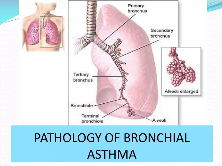
PATHOLOGY OF BRONCHIAL ASTHMA. Bronchial Asthma. Define bronchial asthma. o Describe types of bronchial asthma. o Discuss etiopathogenesis of asthma. o Describe morphological features. Obstructive and restrictive lung diseases. Obstructive lung diseases

E N D
Bronchial Asthma • Define bronchial asthma. • o Describe types of bronchial asthma. • o Discuss etiopathogenesis of asthma. • o Describe morphological features.
Obstructive and restrictive lung diseases • Obstructive lung diseases • (i.e. increased resistance to air flow) include:- • 1. BronchialAsthma. • 2. Emphysema. • 3. Chronic bronchitis. • 4. Bronchiectasis. • 5. Cystic fibrosis and bronchiolitis. • Why should we care about asthma?
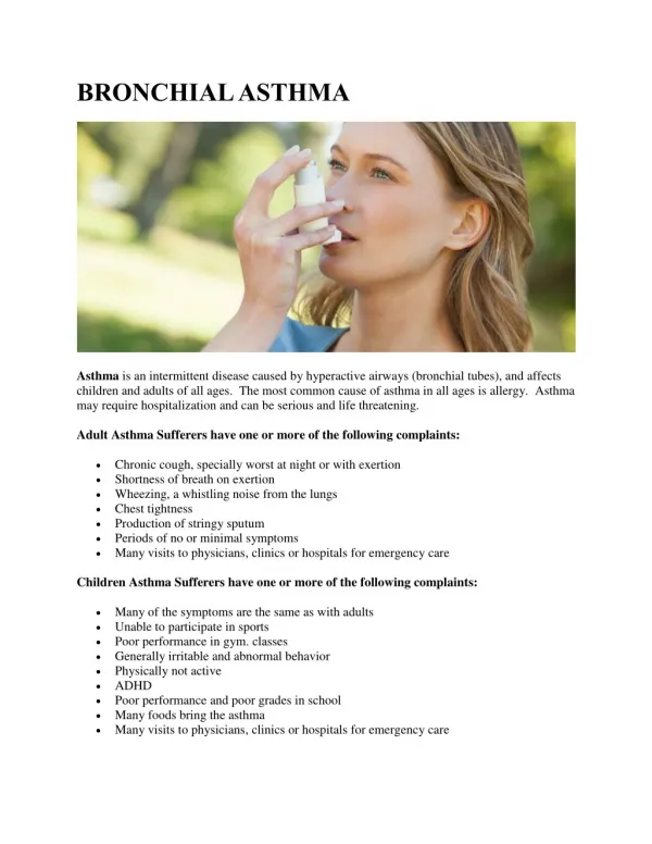
What is Asthma? • Definition: • Asthma is a chronic inflammatory disorder of the airways that causes recurrentspasmodic episodes, due to increased hyperirritability or responsiveness of the bronchial tree to various stimuli. • This associated with these clinical manifestations: • 1. Wheezing. • 2. Breathlessness. • 3. Chest tightness. • 4. Cough, particularly at night and/or in the early morning. • It is manifested physiologically by a widespread narrowing of the air passages, which may be relieved spontaneously or as a result of therapy. Can not cure but can controlled. • Life threatening disease • How long this episodes taking place? (Mild, severe) (Min\hr)
Bronchial asthma risk factors • 1- Atopy(allergic asthma- largest risk factor- genetic). • _______________________________ 2 – Environmental factors( viruses, occupational exposures, allergens, cold air, dust, smoking ,others… ). ____________________________________ 3- family history. ________________________________________________________________________________________________ 4- others……………..
Common triggers for asthma Disease characteristics
Bronchial asthma Classifications Asthma may be categorized into types • ____________________________________________ _______________________________________________ 3- Bronchoconstriction triggering agents - include ( a) Seasonal asthma ( b) Exercise-induced asthma. (c) Drug-induced asthma (e.g., aspirin & NSAID). (d) Occupationalasthma (e) Eemotional asthma . (f ) Asthmaticbronchitis in smokers. _________________________________________ 4-Recent studies added three subphenotypes of Asthma , based on Airway inflammation pattern.
Asthma Types • 1- Atopic asthma (allergicsensitization, Extrinsic) : • Classic example of type I IgE-mediated hypersensitivity reaction. • Usually encountered in patient known case of rhinitis, eczema. • Genetic predisposition. • A positive family history of asthma is common. • Begins in childhood. • Triggered by environmental allergens, such as dusts, pollens, roach or animal dander, and certain types of foods., etc… • Diagnosis: clinical diagnosis is essential +……………………………. • (a) Skin test : Using the offending antigen immediate wheal-and-flare reaction. • (b) Serum radioallergosorbent tests (called RAST): TO identify the presence of IgE specific for a panel of allergens.
Asthma Types 2. Non-atopic asthma: - Non allergic. - Triggered commonly by Respiratory infection due to viruses (e.g., rhinovirus, parainfluenza virus). - Family history: less common. - Skin test: reveals negative reaction. - Mechanism: It is thought thatvirus-induced inflammation of the respiratory mucosa lowers the threshold of the subepithelialvagal receptorsto irritants.
Asthma Types 3- Bronchoconstriction triggering agents • (a) Drug-Induced Asthma. • - Aspirin-sensitive asthma + NSAID occurring with recurrent rhinitis and nasal polyps. • -Others examples: adrenergic antagonists, coloring agents . • Commonly occurs in adult. • Mechanism: • Aspirin inhibiting the cyclooxygenase pathway of arachidonicacid metabolism without affecting the lipoxygenase route, thus tipping the balance toward elaboration of the bronchoconstrictorleukotrienes.
Asthma Types • (b) Occupational Asthma. • Caused or worsened by breathing in irritants on the job. • Triggered\stimulated by: • 1) Fumes (epoxy resins, plastics) • 2) Metal and dusts (platinum, wood, cotton) • 3) Chemicals and Gases (formaldehyde, penicillin products, toluene, enzymes). • 4) Animal substances (5) Plants • - Minute quantities & Repeated exposure. • - Mechanisms: • According to stimulus include:- • Type I hypersensitivity reactions . • Liberation of bronchoconstrictor substances. • Hypersensitivity responses of unknown origin.
Bronchial asthma • 4- Pattern of the Airway inflammation : • 1) Eosinophilic asthma. • 2) Neutrophilic asthma. • 3) Mixed inflammatory asthma. • 4) Pauci-granulocytic asthma. • These subgroups may differ in their: • (a) Etiology. • (b) Immunopathology. • (c ) Response to treatment.
Asthma Pathogenesis-1 • GENETIC CONSIDERATIONS • Genetic predisposition • In case of Atopic asthma- type I hypersensitivity • Inheritance of susceptibility genes (postulation) that makes individuals prone to develop strong TH2 reactions against environmental antigens (allergens)
Asthma Pathogenesis-2 • 1. The airway epithelium and submucosa contain dendritic cells that capture &process antigen \allergens. • Initial sensitization stimulate induction of TH2 cells. • 2. TH2 cells secrete cytokines e.g.(IL-4, IL-5,IL-13) that promote allergic inflammation and stimulate B cells to produce IgE and other antibodies. • 3. Action of Cytokins • (a) IL-4 Production of IgE by B cells. • (b)IL-5 Activates recruited eosinophils. • (c ) IL-13 Mucus secretion(bronchial submucosal glands) also Promotes IgE production by B cells.
Asthma Pathogenesis-3 (Early & Late reaction) • 3. IgE coats submucosal mast cells. • 4. Repeat exposure triggers the mast cells to release granule contents and produce cytokines and other mediators induce the early-phase (immediate hypersensitivity) reaction and the late-phase reaction
Asthma Pathogenesis-4 (Early reaction- Minutes) - AntigensTh2+ IgEproductionIgEbinding to mast cells leads to Eosp. recruitment& release of primary mediators= (Histamine, chemotactic factors, and secondary mediators i.e. leukotriens, prostaglandins, cytokines and neuropeptides). This results in: • (A) Bronchospasm- triggered by direct stimulation of subepithelialvagal (parasympathetic) receptors through both central and local reflexes . • (B) Secretion of mucus. • (C) Variable degree of vasodilatation& increase permeability. • (D) Accumulation of leukocytes.
Asthma Pathogenesis-5 (Late reaction- Hours) • - 6- 10 hr later, produces a continued state of airway hyperresponsiveness with eosinophilic and neutrophilic infiltration. (steroid helpful to treat this stage) Components: consists largely of inflammation with recruitment of leukocytes= ( Eosinophils, neutrophils, and more T cells). - Leukocyte recruitment is stimulated by chemokines produced by mast cells, Epithelial cells (eotaxin ) and T cells, and by other cytokines. - Outcome: persistent bronchospasm, edema, and necrosis of epithelialcells by The major basic protein of eosinophils.
Atopic asthma Epithelial cell injury Allergen IgE Mucus secretion Muscle contraction Mucus secretion Mast cell Muscle contraction Release of inflammatory mediators Recruitment of leukocytes Acute phase Late-phase
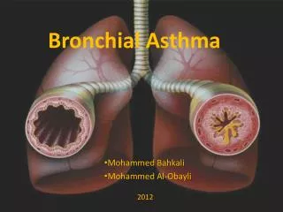
Morphology of Asthma • - Gross appearance: - Done in lungs necropsy-study • 1. Over-inflated and failure to collapse with patchy atelectasis (pt. dying with Status asthmaticus). • 2. Occlusion of airways by gelatinous mucous & exudate • plugs (the bronchial branches up to terminal bronchioles) Microscopic appearance: • Edema, hyperemia, and inflammatory infiltrate (with excess eosinophils) in the bronchial walls. • Increase in the size of submucosal glands (hypertrophy\ hyperplasia) • Patchy necrosis and shedding of epithelial cells. • Over time – features of airway remodeling. -
Airway remodeling. • Overall thickening of airway wall . Reduction of diameter • Basement membrane fibrosis(BM thickening). • Increases muscle mass (Hypertrophy and/or hyperplasia). • Increased in size and number of blood vessels. • Increase number of the submucosal glands. • Mucus metaplasia of epithelium. • Increased fibrogenic factors collagen type I,II “scar” • Irreversible Airflow obstruction.
Epithelial remodeling CONTROL • Epithelium is damaged • New blood vessels • New muscle • New mucosal cells • Collagen deposition
Investigating asthma used to monitor a person's ability to breathe out air. Reduced when airways are constricted. • 1. Elevated eosinophil count in the peripheral blood &CBC. • 2. Sputum smear- Whorled mucous plugs = Curschmann spirals. • 3. Sputum smear- Charcot-Leyden crystals in the sputum (Specially in atopic asthma). (Sputum). • 4. Sputum -Creola bodies (desquamated epithelial cells) • 5. Skin Test • 6. RAST test.