Download
1 / 62
660 likes | 1.14k Vues
Membrane Transport of Small Molecules and the Electrical Properties of Membranes. Chapter 11, Molecular Biology of the Cell Jin-Chung Chen.

E N D
Membrane Transport of Small Molecules and the Electrical Properties of Membranes Chapter 11, Molecular Biology of the Cell Jin-Chung Chen
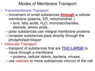
Lipid bilayer: to separate the inner and outer environment of the cell; require special design of carrier protein to transport the water-soluble nutrient into the cell 15-30% membrane proteins exert function of transport (special case: 2/3 of the metabolic energy consumed on transport in special cell) The higher the hydrophobicity or non-polar molecule make ease to pass through the membrane structure Carrier protein disorder: cystinuria-cystine accumulates in the uria formed kidney stone
Two classes of transfer protein: (1) Carrier protein (permease, transporter, pump) : for specific molecule; usually coupled with energy source (2) Channel protein: inorganic ions; down to its concentration gradient; fast overall, transfer proteins create electrical (because of membrane potential) and concentration gradient, in turn, used as a driving force(electrochemical gradient) to facilitate the transport process
Electrochemical gradient combines with membrane potential as a driving force
Involved conformation change Both carrier and channel contain specialized transmembrane domain Transport through channel: fast transport
Passive transport: facilitated diffusion for all channel protein and part of carrier (concentration or electrochemical gradient) Active transport: against electrochemical gradient; mediated by carrier (pump); use ATP or ion gradient as energy source
Ionophores: small hydrophobic molecules (originally formed by microorganism) in membrane to transport specific ions; not coupled to energy source (down to concentration gradient) • Valinomycin: potassium ion (mobile) • FCCP: hydrogen ion (mobile) • A23187: calcium and magnesium ion (mobile) • Gramicidin A: monovalent cation (channel former)
Kinetics of simple diffusion and carrier-mediated diffusion (expressed as Vmax/Km or Bmax/Kd)
Three ways of driving active transport: • coupled carrier: downhill solute drive uphill solute • ATP-driven: ATP hydrolysis to drive uphill solute • light-driven: light energy trigger uphill solute bacteriorhodopsin
Three types of carrier-mediated transport: uniport, symport ( kidney/GI epitghelial cells ) and antiport (determined by its path direction)
Application of Na+-driven carrier proteins (use ion gradient as energy source to drive solute): • Na+ driven glucose into the cell (stomach brush border) (symporter) • Na+ pump that regulates the cellular pH : • Na+-H+ exchanger: influx of Na+ while pump out H+ (maintain the inner pH of the cell) • Na+-driven Cl-HCO3- exchanger: influx Na+ and HCO3- while pump out H+ and Cl- (maintain pH of the cell) • Na+-HCO3- symporter of the glial cell:electrogenic, pump in one Na+ and two HCO3- help to regulate the extracellular pH in near-by neurons during electrical activity
Transcellular transporter: apical to basolateral transport nutrients (intestinal epithelial cells: microvilli)
P-type transporter: 1. Na+-K+ pump • [K+]i: [K+]o = 20 : 1 whiles [Na+]i: [Na+]o = 1: 15 across the cell membrane; • The concentration difference is maintained by Na+-K+ pump (sodium pump; all the animal cells) • The pump is an antiporter, executes active transport and also is an ATPase; its consists of a catalytic subunit (1000 a.a.) and a small glycoprotein (critical for membrane docking) • Most important and energy consumption protein on the membrane • Pump 3 Na+ out in exchange of 2 K+ in (electrogenic)
Immunocytochemical localization of the Na,K pump in choroid plexus. Choroid plexus contains epithelial cells with intensely stained microvillar and intermicrovillar plasma membranes. The basal and lateral plasma membrane surfaces are not stained. Bar = 2 µm.
Schematic diagram of Na-K pump (P-type transport ATPase: can be reversed experimentally to produce ATP
Aspartic acid Na+-dependent phosphorylation; K+-dependent dephosphorylation
Down-regulation of Na,K pumps can be initiated by dopamine via GPCR activation of endocytosis
Na+-K+ pump regulates cell volume • Water move along with the solutes determines the cell volume (osmolarity; tonicity) • Hypotonic vs. hypertonic solution • The fixed anions (nucleic acid and proteins) inside the cell try to pull the water molecule; while excellular Na+ (driven by pump) and Cl- (expelled by membrane potential) ions balance the force • Ouabain: Na-K pump inhibitor; cell swell and burst due to break up the net Na+ outflow
Calcium homeostasis: Cytoplasmic Ca2+ is regulated coordinately by a Na+/Ca2+ antiporter in plasma membranes and by P-type Ca-ATPases in plasma membranes and endoplasmic reticulum.
P-type transporter: 2. Calcium pump (Ca2+-ATPase) • Eucaryotic cells: [Ca2+]i: [Ca2+]o = 10-7 M: 10-3M • Ca2+ pump located in the endoplasmic reticulum (SR in muscle cell consist of 90% of membrane protein) • Ca2+ pump of the SR brings the calcium ion into the SR for storage, upon activation release the calcium into the cytosol for Ca2+-dependent cell signaling • Contains 10 transmembrane -helices; two calcium ion bind to calcium binding domain cause phosphorylation and lead Ca2+ release into the SR
ABC Transporter: transport ATPase • Largest transport ATPase family (> 50 members) • Contains four domains: two hydrophobic domains each with six transmembrane span (function to translocate) and two ATP-binding cassettes (ABC) • In procaryotes, the transporter locates in the inner membrane to carry nutrients into the cell • The counterpart of the eucaryotic: multidrug resistance (MDR) protein, which produce resistance to drug (i.e. chloroquine and anticancer drug resistance) • Cyctic fibrosis: a mutation on one ABC transporter (cyctic fibrosis transmembrane regulator [CFTR] protein) that function as a Cl- channel in the epithelial
Gram-negative bacteria (cassettes): ABC
Ion channels: channels mediate inorganic ion transport • Narrow, highly selective pores that can open and close • Approximately 100 million ions can pass through / second (105 times greater as compared to any carrier) • Can not couple to energy source to perform active transport (always passive – downhill) • They are gated (open and close status); prolong stimulation would desensitized or inactivated ( closed; usually through phorphorylation) • All animal cells contain ion channels, not limited to neuron; each neuron might have more than 10 types of channel
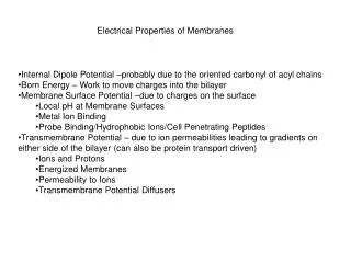
Establish the Membrane Potential • Active electrogenic pump (such as Na-K pump) and passive ion diffusion create the difference of ionic strength across the membrane; hence the difference in electrical charge on two side of the membrane • Major force: Na+-K+ pump and K+ leaking channels (more positive excellularly as compared to negative intracellularly) • a. High concentration of potassium ion inside the cells (due to Na-K pump) keep the electric balance of the negative charged macromolecule • b. K+ leak out down to its concentration gradient • 3. Resting membrane potential: no net flow of ions across the membrane
Number of ions that move across the membrane to set up the membrane potential actually is quite minute
Nernst Equation (and Goldmann equation) RT Co V = zF ln Ci V= the equillibrium potential in volts Co/Ci = outside and inside concentration of the ion, respectively R= the gas constant (2 cal mol-1K-1) T= the absolute temperature (K) F= Faraday’s constant (2.3 x 10⁴ cal V-1 mol-1) z= the valence (charge) of the ion ln= logarithm to the base e
The more permeable the membrane for a given ion, the more strongly the membrane potential tends to be driven toward the equilibrum value for that ion: changes in a membrane’s permeability to ions can cause significant changes in the membrane potential ( resting membrane potential will be determined by most permeable ion(s) )
Diagram of the functional units of a voltage-gated Na+ channel, the hypothesized binding sites for several drugs and toxins are illustrated to affect the opening and closing of the channels
Transmembrane organization of voltage-gated Ca2+ channels, K+ channels and relatives.
3-D Structure of Potassium Channel (exhibited only 2 out of 4 transmembrane subunits) Separated about 8 A
Selection of ion by selectivity filter Sodium ion is too small for carbonyl oxygen interaction and water expel
The ion selectivity filter and pore of Na+ and K+ channel illustrated with the extracellular side upwards. The four ion-coordination sites are thought to work in pairs. In one cycle of outward K+ conductance, K+ ions occupy sites 1 and 3 (orange), shift to sites 2 and 4 (gray), and then the K+ ion in site 4 moves into the extracellular space while a new K+ ion occupies site 1 and the K+ ion in site 2 moves to site 3, re-establishing the initial state.
Generation of an Action Potential • An electrical stimulus that exceeds a certain threshold triggers an electrical activity that is (a) self-propagated and (b) sustained by automatic amplification • Voltage-gated cation channels are responsible for generating the action potential • Consequence of an action potential: • a. stimulus triggers voltage-gated Na+ channel to open • b. more Na+ channel open in neighboring area, cause depolarization (reach Na+ equilibrum potential [+50mV]) • c. Na+ channel inactivated • d. voltage-gated K+ channel open (bring membrane potential to resting status; reach K+ equilibrium potential [-70mv])
N-terminal 20 a.a. function as a tethered ball to inactivate the potassium channel after activation
A hinged-lid model for Na+ channel inactivation, illustrating the inactivation gate formed by the intracellular segment connecting domains III and IV and the critical cluster of hydrophobic residues that forms a latch holding the inactivation gate closed.
Function of Myelination: insulated neurons to increase the rate of propagation; formed by Schwann cells (peripheral) or oligodendrocytes (central) Node of Ranvier: myelin sheath space; most Na+ channels concentrated here Saltatory conduction: action potential propogate node to node (travel faster and metabolic energy is conserved)
Patch clamp technique : allow current recording from single channel
Individual volgate-gated Na+ channel open in all-or-none fashion; the aggregate curent (approached by intracellular recording) across the membrane of entire cell represent the total number of channels ( not the degree) that open at a given time Voltage-gated cation channels (Na+, K+ or Ca2+ ) are evolutionarily and structurally related
Neurotransmission from presynaptic to postsynaptic Transmitter-gated ion channel is not sensitive to membrane potential (can not produce self-excitation). They produce local permeability changes (depend on numbers of released transmitter) until can open nearby voltage-gated cation channels to initiate an action potential of the next neuron Re-uptake Cl--dependent transporter
Neurotransmitter can determine post-synaptic excitation or inhibition (channel receptor) • Excitatory neurotransmitter: open cation channel (Ca2+, Na+) (i.e. NMDA glutamate receptor) • Inhibitory neurotransmitter: open Cl- or K+ channel (i.e. GABAA receptor)