Download
1 / 58
620 likes | 898 Vues
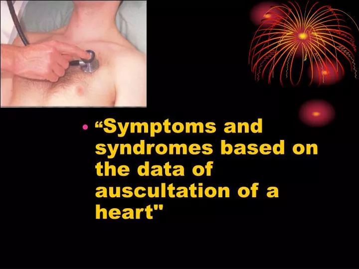
“ Symptoms and syndromes based on the data of auscultation of a heart ". Auscultation was inculcated by French physitian Rene Laennec. Рис. 10. Стетоскопи тверд!. First device for auscultation was a stetoscope. First binaural stetoscope. First phonendoscope. Modern stetophonendoscope.

E N D
“Symptoms and syndromes based on the data of auscultation of a heart"
Auscultation was inculcated by French physitian Rene Laennec Рис. 10. Стетоскопи тверд!.
Sounds heard by stetoscope is called heart sounds. They are created due to vibrations of heart structures during their functioning
Formation of heart soundsa—atrial component (heard sometimes as an independent fourth sound); b—valvular component of the first sound; c—muscular component of the first sound; d—vascular component of the first sound; e—formation of the second sound; /—formation of the third sound
Auscultation involves listening for heart sounds with the stethoscope, similar to the procedure used in assessing breath sounds • The sounds produced by a working heart are called heart sounds. Two sounds can be well heard in a healthy subject; the first sound, which is produced during systole and the second sound, which occurs during diastole.
Сomponents of heart sounds • I heart sound: • the valve component, i.e. vibrations of the cusps of the atrioventricular valves during the isometric contraction phase • the muscular one due to the myocardial isometric contraction • the vascular one. This is due to vibrations of the nearest portions of the aorta and the pulmonary trunk caused by their distention with the blood during the ejection phase • Atrial one is generated by vibrations caused by atrial contractions • II heart sound: • The second sound is generated by vibrations arising at the early diastole when the semilunar cusps of the aortic valve and the pulmonary trunk are shut (the valve component) and by vibration of the walls at the point of origination of these vessels (the vascular component). • The intensity of myocardial and valvular vibrations depends on the rate of ventricular contractions: the higher the rate of their contractions and the faster the intraventricular pressure grows, the greater is the intensity of these vibrations.
Sequence of auscultation • The mitral valve - at the heart apex; • the aortic valve - in the second intercostal space to the right of the sternum), • the pulmonary valve - in the second intercostal space, to the left of the sternum, • tricuspid valve - at the base of the xyphoid process, • the aortic valve again at the Botkin-Erb point.
Differential features of I and II heart sounds I heart sound II heart sound The place of best hearing Heart apex Heart basis Relation to cardiac circle After the longer pause After the shorter pause Duration 0,09-0,12 sec 0,05-0,07 sec Relation to the carotid pulsation Coincides Doesn’t coincide Relation to the apex beat Coincides Doesn’t coincide
For differentiation of I and II heart sounds in tachycardia it is necessary to check which of them is synchronous with carotic artery pulsation
Intensity of the heart sounds may depend on conditions of the sound wave transmission • The intensity of both heart sounds decreases if their transmission to the chest becomes difficult: • subcutaneous fat or muscles of the chest are overdeveloped, • lung emphysema, • liquid in the left pleural cavity, • other affections that separate the heart from the anterior chest wall. • If conditions for sound transmission are improved in decreased myocardial contractility: • in myocarditis, • myocardial dystrophy, • cardiosclerosis, • collapse, • accumulation of fluid in the pericardial cavity.
The intensity of the heart sounds increases if their transmission to the chest becomes better: • thin chest wall, • the lung edges are sclerosed, • the heart is pressed against the anterior chest wall by a growing tumour in the posterior mediastinum, • by the resonance in large empty cavities filled with air (a large cavern in the lung, large gastric air-bubble). • if the blood viscosity decreases (in anaemia) or left ventricular feeling drops (bleeding). due to the effect of the sympathetic nervous system on the heart: • in physical and emotional strain, • during exercise, • in patients toxic goitre.
Scheme of weakening and intensification of both heart sounds
Separate changes of one heart sound (I or II): • First heart sound diminishes: • in the mitral and aortic valve insufficiency (at the apex). In tricuspid and pulmonary valve failure, the diminution of the first heart sound will be better heard at the base of the xiphoid process, • at the heart apex in stenotic aortal orifice, • In diffuse affections of the myocardium (due to dystrophy, cardiosclerosis or myocarditis), the first heart sound only may be diminished because its muscular component also diminishes in these cases. • The first sound increases at the heart apex if the left ventricle is not adequately filled with blood during diastole: • in stenosis of the left atrioventricular orifice, • In extrasystole. • Thesecondsound can be inaudible over the aorta if: • the aortic valve is much destroyed, • diminishes over the aorta in cases with marked hypotension; • diminishes over the pulmonary trunk in cases with aortic valve incompetence (in very rare cases), • in decreased pressure in the lesser circulation. • The second sound may increase either over the aorta or over the pulmonary trunk indicating hypertension in the proper circle of circulation.
Splitting or reduplication of the sounds occurs in asynchronous workand right chambers of the heart • Asynchronous closure of the right- and left ventricular valves splits the first sound while asynchronous closure of thesemilunar valves causes reduplication of the second heart sound. • Reduplication or splitting of the first soundis due to asynchronous closure of the atrioventricular valves, e.g. during very deep expiration, when the blood is ejected into the left atrium with a greater force to prevent the closure of the mitral valve; • Pathological reduplication of the first sound can occur in impaired intraventricular conduction (through the His bundle) as a result of delays systole of one of the ventricles.
The second sound is reduplicated more frequently • due to asynchronous closure ofthe aorta and pulmonary trunk valve • diminished or increased filling of one of the ventricles or when pressure in the aorta or the pulmonary artery changes. • Physiological reduplication is mostly connected with various respiratory phases • Pathological reduplication of the second sound can be due to delayed closure of the aortic valve in persons suffering from essential hypertension, or if the closure of the pulmonary valve is delayed at increased pressure in the lesser circulation
Adventitious heart sounds • The third heart sound (S3) is the result of vibrations produced during ventricular filling. It is normally heard only in some children and young adults, but it is considered abnormal in older individuals. It arises in 0.15—1.12 s from the beginning of the second sound. • The forth heart sound (S4) is caused by the recoil of vibrations between the atria and ventricles following atrial contraction, at the end of diastole. It is rarely heard as a normal heart sound; usually it is considered indicative of further cardiac evaluation. • Both S3 and S4 may be recorded in heart failure indicating poor muscular tone of the left ventricle. • The mitral valve opening sound (opening snup) is heard at the heart apex of patients with mitral stenosis 0.07-0.13 s following the second sound, during diastole. • Extra-pericardial-sound can occur in pericardial adhesion. It originates during diastole, 0.08-0.14 s after the second sound, and is generated by the vibrating pericardium during the rapid dilatation of the ventricles at the beginning of diastole.
Heart melodies • gallop rhythm • The gallop rrhythm is conditionally divides into protodiastolic, mesodiastolic and presystolic. • is better auscultated directly by earin the apical region at left lateral recumbent position of the patient, in III- IV intercostal spaes to the left.
Triple rrhythm (Rhithmus coeturnici) • It is a cardiac rhythm which is auscultated only in mitral stenosis and arises if there is presence of such an adventitious sound as mitral click (or sound of opening of mitral valve) together with slapping first and second sounds.
Pendulum rhythm • In the case of pendulum rhythm the large (diastolic) heart pause is so shortened, that becomes an equal to small (systolic) pause. The sound phenomenon, which one arises thus, reminds of even pendulum swinging. Such rhythm disturbance meets usually at heavy lesions of heart muscle . If pendulum rhythm is accompaning by sharp heart acceleration, this phenomenon is called as embriocardia.
Cardiac murmurs-phenpmena which arise due to pathological blood flow in the heart • Intracardial murmurs: • Organic and functional (relative), • Systolic and diastolic, • Ejection and regurgitation murmurs, • They are also different in character, intensity, duration. • Extracardial (pericarial friction murmur and pleuropericardial murmur)
Properties of murmurs • Duration • Character and pitch • Location • Radiation • Intensity
Grades of intensity of murmur • Heard by an expert in optimum conditions • Heard by a non-expert in optimum conditions • Easily heard; no thrill • A loud murmur, with a thrill • Very loud, often heard over wide area, with thrill • Extremely loud, heard without stethoscope
Causes of systolic murmurs • Ejection systolic murmur • Increased flow through normal valves • • 'Innocent systolic murmur': • fever • athletes (bradycardia -> large stroke volume) • pregnancy (cardiac output maximum at 15 weeks) • Atrial septal defect (pulmonary flow murmur) • Severe anaemia • Normal or reduced flow though stenotic valve • Aortic stenosis • Pulmonary stenosis • Other causes of flow murmurs • Hypertrophic obstructive cardiomyopathy (obstruction at subvalvular level) • Aortic regurgitation (aortic flow murmur) • Pansystolic murmurs • I caused by a systolic leak from a high to a lower pressure chamber Mitral regurgitation Tricuspid regurgitation Ventricular septal defect Leaking mitral or tricuspid prosthesis • or bradycardia. Atrial septal defect is characterized
At an auscultation it is necessary to determine: • 1) relation of murmur to the phase of cardiac cycle (systole or diastole); • 2) properties of murmur, its character, intensity, duration; • 3) localization of murmur, i.e. place of the best auscultation; • 4) condution of murmur (irradiation).
Differentiation of functional and organic murmurs • in the most cases functional murmurs are systolic; • the murmurs are changeable, can arise and decrease in intensity or even disappear at various positions of a body, after an exercise, stress, in different phases of respiration; • most often they are auscultated above a pulmonary trunk, less often — above heart apex, • the murmurs are short, seldom occupy all systole; mild and blowing in character; • the murmurs are usually auscultated on a circumscribed field and are not conducted far from the place of occurence; • The functional murmurs are not accompanied by other attributes of valvular lesions (enlargement of heart chambers, change of sounds etc.).
The pericardial friction • It is develops in change of visceral and parietal pericardiac layers, when the fibrin (is postponed at a pericarditis), or cancerous nodules are deposied on them. • Differential features: • It is heart equally over the whole heart area, • It intensifies if to press motightly to the heart area with a phonendoscope and at inclination of a trunk forward, • It is sinchronous with heart contractions (is heart in systole and diastole), • it is changeable, disappear and appear again.