Download
1 / 207
2.07k likes | 2.22k Vues
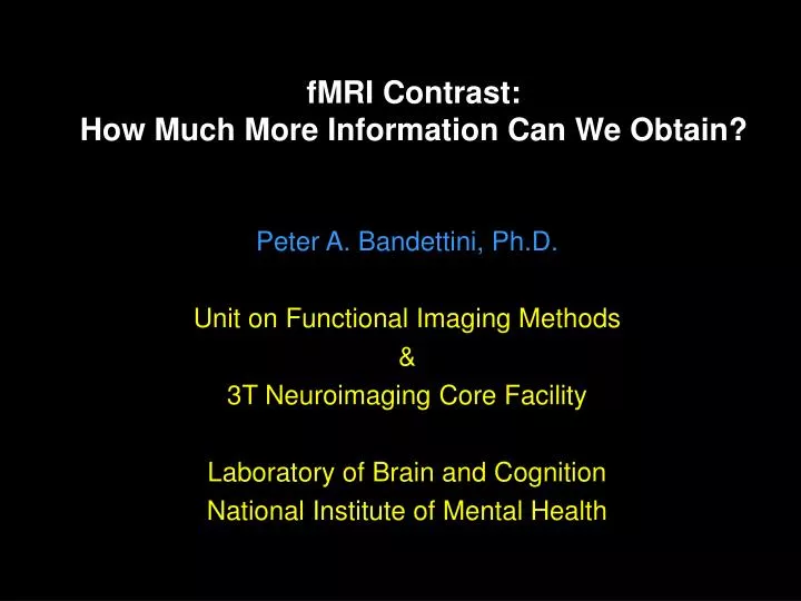
fMRI Contrast: How Much More Information Can We Obtain?. Peter A. Bandettini, Ph.D. Unit on Functional Imaging Methods & 3T Neuroimaging Core Facility Laboratory of Brain and Cognition National Institute of Mental Health. BOLD Contrast. Neuronal. Measured. Activation. fMRI. Signal. ?.

E N D
fMRI Contrast: How Much More Information Can We Obtain? Peter A. Bandettini, Ph.D. Unit on Functional Imaging Methods & 3T Neuroimaging Core Facility Laboratory of Brain and Cognition National Institute of Mental Health
Neuronal Measured Activation fMRI Signal ? ? Hemodynamics The continuing challenge is to make progressively more precise neuronal, metabolic, and hemodynamic inferences across spatial and temporal scales.
Pulse Sequence Modulation Task Modulation & Measurement Neuronal Measured Activation fMRI Signal ? ? Hemodynamics Physiologic Modulation / Measurement
1991- 2 • TE dependence • Field Strength Dependence • Resolution Dependence • Pulse sequence dependence (T2 and T2*) • Dynamics: latency and return to baseline • First BOLD models • Correlation of BOLD with parametric task manipulation • Post undershoot • NMR phase shift observation • Angio and venogram correlation • Effects of Physiologic Stress • Retinotopy • Cognitive mapping • Event - related fMRI • Parametric task design • Sub-millimeter resolution fMRI • Diffusion weighting dependence (IV contribution) • Physiologic fluctuations • Extended duration stimulation effects • Pre undershoot • Flow and BOLD comparisons (dynamics and magnitude) • Blood volume dynamics • Simultaneous flow and BOLD acquisition • Ocular Dominance Column Mapping • CMRO2 Mapping • Randomized ER-fMRI task design • Balloon Model • Baseline Blood Oxygenation Quantitation • Mental Chronomitry • Linearity of BOLD signal change BOLD Contrast Advancements 1992- 3 1993- 4 1994 - 5 1995 - 6 1996 - 7 1997- 8 1998 - 9 1999 - 2000
Pulse Sequence Modulation Task Modulation & Measurement Neuronal Measured Activation fMRI Signal ? ? Hemodynamics Physiologic Modulation / Measurement
+ 2 sec Latency - 2 sec Magnitude
9.0 seconds 15 seconds 500 msec 500 msec 20 30 10 Time (seconds)
Regions of Interest Used for Hemi-Field Experiment Left Hemisphere Right Hemisphere
3.2 2.4 1.6 0.8 0 -0.8 -1.6 -2.4 0 10 20 30 Hemi-field with 500 msec asynchrony Average of 6 runs Standard Deviations Shown Percent MR Signal Strength Time (seconds)
500 ms 500 ms RightHemifield Left Hemifield + 2.5 s - = 0 s - 2.5 s
Motor Cortex Auditory Cortex
Methods Motor Visual Stimulus Duration (SD) … SD = 500 ms SD = 250 ms … SD = 1000 ms SD = 500 ms … SD = 2000 ms SD = 1000 ms … SD = 4000 ms SD = 2000 ms 16 s … Blocked Trial 20 s
20 s 2000 ms 1000 ms 500 ms 250 ms 0 10 20 30 40 0 10 20 30 40 BOLD response is nonlinear Observed response Linear response Short duration stimuli produce larger responses than expected
a t Compute nonlinearity (for each voxel) • Amplitude of Response Fit ideal (linear) to response • Area under response / Stimulus Duration Output Area / Input Area
6 6 4 f (SD) 4 f (SD) 2 2 0 1 2 3 4 5 0 1 2 3 4 5 Stimulus Duration Stimulus Duration 6 5 4 4 3 2 2 1 0 1 2 3 4 5 0 1 2 3 4 5 Nonlinearity Visual Motor Magnitude linear Area Output / input Output / input Stimulus Duration Stimulus Duration
Measure of Nonlinearity • Area under nonlinearity curve f (SD) Area 1 0 1 2 3 4 5 Stimulus Duration (s) • Slope of nonlinearity curve • difference at each SD
Results – visual task Nonlinearity Magnitude Latency
8 f (SD) 6 4 2 0 10 20 30 40 0 1 2 3 4 5 0 10 20 30 40 Stimulus Duration -2 8 f (SD) 6 60 4 40 2 20 0 1 2 3 4 5 0 2 0 2 4 6 8 Stimulus Duration -2 nonlinearity Results – visual task
Results – motor task Nonlinearity Magnitude Latency
8 f (SD) 6 4 2 60 0 10 20 30 40 0 10 20 30 40 40 0 1 2 3 4 5 Stimulus Duration 20 0 2 0 2 4 6 8 nonlinearity Results – motor task 8 f (SD) 6 4 2 0 1 2 3 4 5 Stimulus Duration
Reproducibility Visual task Motor task Nonlinearity1 Nonlinearity1 Nonlinearity2 Nonlinearity2 Experiment 1 Experiment 2 Experiment 1 Experiment 2
Conclusions • Responses to short duration stimuli are larger than predicted from a linear system • Spatial variation in this nonlinear relationship is seen • The variation in nonlinearity is not significantly correlated with magnitude or latency
Pulse Sequence Modulation Task Modulation & Measurement Neuronal Measured Activation fMRI Signal ? ? Hemodynamics Physiologic Modulation / Measurement
Finger Movement Anatomical 12% O2 5% CO2
% =
Pulse Sequence Modulation Task Modulation & Measurement Neuronal Measured Activation fMRI Signal ? ? Hemodynamics Physiologic Modulation / Measurement
Activation-induced MR Signal Change Mechanisms T1 Inflow T2 T2*
T1 - weighted T2* weighted T1 and T2* weighted
Perfusion Rest Activation BOLD
Anatomy BOLD Perfusion
b = 0 b = 10 b = 50 b = 160
= 6 =12 = 24 = 48 Wright et al. JMRI, 1: 275-228, 1991
D = 1 µm2 / ms -> 2.5 µm2 / ms = 6 =12 = 24 = 48
Field -Strength Dependence of T2* and T2 Hct = 44,= 48, %HbO2 = 60, T2o = 250 ms, T2’ = 120 ms
3T Spin-Echo TE = 105 ms TR = ∞ Gradient-Echo TE = 50 ms Gradient-Echo functional TE = 50 ms Spin-Echo functional TE = 105 ms
Spin echo vs. Gradient echo compartment radius: < 3 µm 3 to 15 µm > 15 µm
∆R2* / ∆R2 1.5 1.5 intravascular 1.5 to 3 extravascular 1.5 intravascular 3 to ∞ extravascular average ∆R2* / ∆R2 ≈ 3 to 4
Spin - Echo Gradient - Echo During Activation Increase Post Activation Undershoot