Download
1 / 46
540 likes | 1.16k Vues
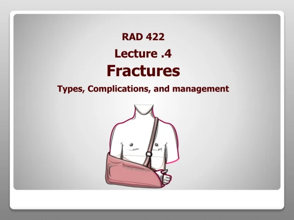
Fractures and Fracture Management. Resident Orientation Course 2012 Presented By Dr. Shinouri. Outline. Definition of Fracture Radiographic and clinical description of Fractures Classification of Fractures Treatment of Fractures. Definition of Fracture.

E N D
Fractures and Fracture Management Resident Orientation Course 2012 Presented By Dr. Shinouri
Outline • Definition of Fracture • Radiographic and clinical description of Fractures • Classification of Fractures • Treatment of Fractures
Definition of Fracture • Disruption in the integrity of living bone, involving injury to the bone marrow, periosteum and adjacent soft tissues • Described radiographically and clinically
Radiograph and Clinical Descriptions • Anatomy: Described in relation to the bones involved and the location within the bone (diaphysis, metaphysis, physis, epiphysis) • Articular surface involvement: Does the fracture have intra-articular involvement?
Anatomy and Articular Surface Epiphysis • Which bone? • Thirds (long bones) • Proximal, middle, distal third • Anatomic orientation • Proximal, distal, medial, lateral, anterior, posterior • Anatomic landmarks • Head, neck, body / shaft, base, condyle • Segment (long bones) • Epiphysis, physis, metaphysis, diaphysis Physis Metaphysis Diaphysis (Shaft) Articular Surface
Radiograph and Clinical Descriptions • Displacement: Is the distal fracture fragment displaced compared with proximal fragment? To what degree is the fracture displaced? • Angulation: The angular deformity is defined in degrees in terms of the distal fragment in relation to the proximal fragment
Radiograph and Clinical Descriptions • Shortening: Has the fracture caused shortening of the involved bone? • Rotation: Described both radiographically and clinically
Radiograph and Clinical Descriptions • Fragmentation • Multi-fragmentary fracture: several breaks in bone creating 2 fragments • Wedge fracture: either spiral (low energy) or bending (high energy) and allow the proximal and distal fracture fragments to contact each other • Simple fractures: spiral, oblique or transverse
Radiograph and Clinical Descriptions • Soft tissue involvement: • Is the fracture open or closed? • Is associated neurologic or vascular injury present? • Is there muscle damage or compartment syndrome?
Closed Fracture • Fracture is not exposed to the environment • All fractures have some degree of soft tissue injury • Commonly classified according to the Tscherne classification
Closed Fracture Classification • Tscherne Classification: • Grade 0: Minimal soft tissue injury; Indirect injury • Grade 2: Direct injury; more extensive soft tissue injury and severe bone injury • Grade 3: Severe injury to soft tissue; degloving with destruction of subcutaneous injury (Compartment syndrome is a concern)
www.orthoinfo.aaos.org Orthopedic Trauma Association
Open Fracture • A break in the skin and underlying soft tissue leading directing into or communicating with the fracture and its hematoma • Serious injury as infection can gain entrance into the body through the wound and endanger limb or life • Described by the Gustilo classification system
Orthopedic Trauma Association www.jbjs.org
Classification of Open Fractures • Type 1: Wound is smaller < 1 cm, clean and caused by fracture fragment that pierces the skin (Low energy injury) • Type II: Wound > 1cm, not contaminated. And without major soft tissue damage defect (Low energy injury)
Classification of Open Fractures • Type III: Wound > 1 cm, with significant soft tissue disruption (High energy trauma) severely unstable fracture with varying degrees of fragmentation • Type III fractures are subdivided: • IIIA: Wound has sufficient soft tissue to cover the bone without the need for local or distant flap coverage
Classification of Open Fractures • Type III fractures: • IIIB: Disruption of soft tissue is extensive, such that local or distant flap coverage is necessary • Wound is often contaminated • IIIC: Associated with an arterial injury that requires repair
Epidemiology • Frequency: • Trauma is the leading cause of death ages 1-34 yrs • Causes more years of lost productivity before age 65 than coronary artery disease, cancer, and stroke combined • Facture incidence is multi-factorial such as age, sex, co-morbidities, lifestyle and occupation (In the US, 5.6 million fractures/year)
Etiology of Fractures • Occur when the force applied to a bone exceeds the strength of the involved bone • Both intrinsic + extrinsic factors are important • Extrinsic factors: include the rate at which the bone’s mechanical load is imposed and the duration, direction, and magnitude of the forces acting on the bone • Intrinsic factors: include the involved bone’s energy-absorbing capacity, modulus of elasticity, fatigue, strength, and density
Etiology of Fractures • Result of direct or indirect trauma • Direct trauma consists of direct force applied to the bone • Direct mechanisms include tapping fractures (eg, bumper injury), penetrating fractures (eg, gunshot wound),and crush fractures • Indirect trauma involves forces acting at a distance from the fracture site such as tension (traction), compressive, and rotational force
Pathophysiology • 5 phases of fracture healing: • Fracture and inflammatory phase • Granulation tissue formation • Callus formation • Lamellar bone deposition • Remodeling
Fracture and Inflammatory Phase • Actual fracture injuries to the bone include insult to the bone marrow, periosteum, and local soft tissues • The most important stage in fracture healing is the inflammatory phase and subsequent hematoma formation • It is during this stage that the cellular signaling mechanisms work through chemotaxis and an inflammatory mechanism to attract the cells necessary to initiate the healing response
Granulation Tissue Formation • Within 7 days, the body forms granulation tissue between the fracture fragments • Various biochemical signaling substances are involved in the formation of the granulation tissue stage, which lasts approximately 2 weeks
Callus Formation • During callus formation, cell proliferation and differentiation begin to produce osteoblasts and chondroblasts in the granulation tissue • The osteoblasts and chondroblasts synthesize the extracellular organic matrices of woven bone and cartilage, and then the newly formed bone is mineralized • This stage requires 4-16 weeks.
Lamellar bone Formation and Remodeling • During the fourth stage: Meshlike callus of woven bone is replaced by lamellar bone, which is organized parallel to the axis of the bone • Final stage: remodeling of the bone at the site of the healing fracture by various cellular types such as osteoclasts • The final 2 stages require 1-4 years
Presentation of Single Limb Injury • Obtain through hisotiry for the mechanism of injury • Obtain history of any previous injury or fracture is mandatory • Obtain complete past medical and surgical history • Including medications and allergies, as well as a social (smoking and illicit drug use) and occupational history
Physical Examination in Single Limb Injury • Physical exam must include a thorough inspection of the integument (with documentation) • If the fracture is open, a clinical photograph may be taken for documentation purposes • Distal neurologic and vascular status must be assessed and documented
Physical Examination in Single Limb Injury • Palpate the entire limb including the joints above + below the injury -- check for areas of pain, effusions, and crepitus • Often, other associated injuries may be present (eg, injuries to the spine with a jumping mechanism of injury) • Assessment of range of motion (ROM) may not be possible, but this should be documented • Assessments for ligamentous injury and tendon rupture
Multiple Traumatic Injuries • Initial assessment of a patient with polytrauma follows the advanced trauma life support (ATLS) protocols • Includes the identification and treatment of life-threatening injuries • The first step is evaluation of the individual's airway, breathing, and circulation • Immediate endotracheal intubation and rapid administration of intravenous fluids may be necessary
Multiple Traumatic Injuries • Spinal precautions must be maintained until injury to the spine can be excluded clinically and radiographically • Radiographs or computed tomography [CT] scans • Once patient is hemodynamically stable • The secondary survey, a complete systems-based physical examination, is performed
Initial Management of Fractures • Consists of realignment of the broken limb segment and then immobilizing the fractured extremity in a splint • Distal neurologic + vascular status must be clinically assessed and documented before and after realignment and splinting
Initial Management of Fractures • If a patient sustains an open fracture, achieving hemostasis as rapidly as possible at the injury site is essential • This is achieved by placing a sterile pressure dressing over the injury • Splinting is critical in providing symptomatic relief for the patient, preventing potential neurologic + vascular injury and further injury to the local soft tissues • Patients should receive adequate analgesics in the form of acetaminophen or opiates, if necessary.
Management of Open Fractures • Treatment goals for open fractures are to: • Prevent infection, to allow the fracture to heal, and to restore function in the injured limb • Once initial assessment, evaluation, and management of any life-threatening injuries are complete, the open fracture is treated • Hemostasis should be obtained, followed by antibiotic administration and tetanus vaccination
Management of Open Fractures • Cefazolin or clindamycin is adequate for type I + type II fractures • Aminoglycoside (eg, gentamicin) can be added for a severely contaminated wound (type III) • If injury is a "barnyard injury" or water-type injury, penicillin should also be added to provide prophylaxis against Clostridium perfringens
Management of Open Fractures • Urgent irrigation and debridement (I&D) of the wound in the operating room is mandatory • For type II and type III injuries, serial I&Ds are recommended every 24-48 hours after the initial debridement until a clean surgical wound is ensured • The wound is closed when it is clean and antibiotics are generally continued until 2 days after the final I&D
Management of Open Fractures • Management of the open fracture depends on the site of injury and type of open fracture • Wound is subsequently stabilized either temporarily or definitively • If soft-tissue coverage over the injury is inadequate • Soft-tissue transfers or free flaps are performed when the wound is clean and the fracture is definitively treated
Fracture Management • Divided into non-operative and operative techniques • Non-operative technique: consists of closed reduction if required followed by a period of immobilization with casting or splinting • Closed reduction is needed if the fracture is significantly displaced or angulated • If the fracture cannot be reduced, surgical intervention may be required
Fracture Management – Surgical Indications • Failed nonoperative (closed) management • Unstable fractures that can’t be maintained in a reduced position • Displaced intra-articular fractures (>2 mm) • Fractures that are known to heal poorly following non- operative management (eg, femoral neck fractures)
Fracture Management – Surgical Indications • Large avulsion fractures that disrupt the muscle-tendon or ligamentous function of an affected joint • Example patella fracture • Impending pathologic fractures • Multiple traumatic injuries involving pelvis, femur, or vertebrae • Unstable open fractures or complicated open fractures
Fracture Management – Surgical Indications • Individuals who are poor candidates for non-operative management that requires prolonged immobilization • Example: elderly patients with proximal femur fractures • Fractures in skeletally immature individuals that have increased risk for growth arrest • Example: Salter-Harris types III-V) • Nonunions or malunions that have failed to respond to non-operative treatment
Contraindications • Active infection (local or systemic) or osteomyelitis • Soft tissues that compromise the overlying fracture or the surgical approach • Medical conditions that contraindicate surgery or anesthesia • Example: recent myocardial infarction • Cases in which amputation would better serve the limb and the patient
References • Buckley, R. General Principles of Fracture Care. Medscape Jan 2010. • Orthopedic Trauma Association.www.ota.org