Download
1 / 58
700 likes | 2.67k Vues
Management of Extremity Fractures. Bucky Boaz, ARNP-C. Upper Extremity Fractures. Commonly encountered in Family Practice Ranked 14 th out of top 20 diagnoses 6% to 15% of orthopedic problems encountered in Family Practice

E N D
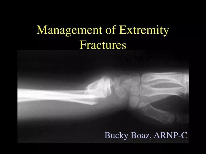
Management of Extremity Fractures Bucky Boaz, ARNP-C
Upper Extremity Fractures • Commonly encountered in Family Practice • Ranked 14th out of top 20 diagnoses • 6% to 15% of orthopedic problems encountered in Family Practice • Most common injuries are fractures of fingers, radius, metacarpals, toes, and fibula • Many can be managed by Family Practice
Metacarpal Fractures • Second most common fracture in primary care • Classified according to location: • Head • Neck • Shaft • Base
Metacarpal Fractures • Most fractures of MC head are comminuted and need ortho referral • Acute mgt: • Immobilize in ulnar or radial gutter splint • Ice • Elevation • Analgesia • Ortho evaluation within 1 week of injury
Metacarpal Fractures • Fractures of MC neck result from direct impact (punching) • Boxer’s Fracture • Head of MC is displaced volarly • Tenderness and swelling over dorsum of hand • Possible pseudocrawling • Hyperextension at MCP and flexion at PIP • Dorsal angulation > 40º
Metacarpal Fractures • Radiographs AP, lat and oblique views • Degree of angulation on lateral view • Expected 15º • Subtract from visualized angulation • More distal greater allowed • Deformity better tolerated in 4th or 5th digits
Ulnar Gutter Splint Metacarpal Fractures • Management • Splint: • MCP 70º to 90º of flexion • Use radial or ulnar gutter • Reduction: • Pseudocrawling • 4th MC > 30º • 5th MC > 40º • May not improve outcome
Metacarpal Fractures • Reduction • Hematoma or ulnar nerve block • 90-90 method: • MCP, PIP, DIP joints flexed 90º • Volar-directed pressure over fracture site • Immob with wrist extension 30º and MCP flexed to 90º
Fracture 4th metacarpal Metacarpal Fractures • Nondisplaced fractures of 2nd and 3rd MCs follow up x-ray within 4-5 days • Fractures to 4th or 5th MCs follow up x-ray 7-10 days • Any change, ortho referral • No contact sports for 4-6 weeks after immobilization
Wrist Anatomy • Metacarpals and phalanges • Trapezium • Carples • Scaphoid (navicular) • Distal radius • Lunate • Triquetrium • Pisiform • Capitate • Hamate • Trapezoid
Wrist Anatomy Dorsal Anatomic Landmarks • Radial styloid • Extensor pollicis brevis • Anatomic snuffbox • Extensor pollicis longus • Lister’s tubercle • Dorsal wrist depression • Ulnar styloid
Wrist Anatomy Volar anatomic landmarks • Radial styloid • Scaphoid tubercle • Carpal tunnel • Hamulus • Pisiform
Scaphoid Fracture • Most commonly fractured carpal bone • 70% - 80% of all carpal bone injuries • 8% of all sports-related fractures • Spans both carpal rows • Susceptible to injury when stress applied to dorsiflexed wrist
Scaphoid Fracture • Patients will complain of wrist pain • Particularly over anatomic snuff box • Swelling • Motion is commonly limited
Scaphoid Fracture • Radiographs need to include scaphoid view • Elongates the scaphoid along its long axis • At least 10%-20% false negative on x-ray
Scaphoid Fracture • Anatomical Importance: • Blood supply from a branch of radial artery enters the distal pole • Retrograde blood flow • Fractures at risk of nonunion or AVN • Proximal • Oblique • displaced
Scaphoid Fracture • Examination • Anatomic snuff box swelling or pain on palpation • Pronation and ulnar deviation exacerbates pain • Axial loading exacerbates pain • Pronation/supination against resistance exacerbates pain (supination more specific)
Scaphoid Fracture • Management • Immobilize even if x-rays negative if warranted • Immobilization with thumb spica • Ortho referral
Colles’ Fracture • Most common fracture of the distal radius • Results from a fall on an outstretched hand (FOOSH) • Dorsal swelling • Eccymosis • “Silver fork” deformity of the hand and wrist
Colles’ Fracture • Radiographs • (AP, lat, & oblique) • Apex volar fracture with dorsal comminution and shortening of the radius • Typically occurs within 2cm of distal radius articular surface
Colles’ Fracture • Definitive care may be provided by primary care provider • Reduction of fracture • Splinting • Ortho referral • Inter-articular fracture needs ortho follow up
Smith’s Fracture • Less common fracture of distal radius • Unstable fracture • Distal fragment is displaced volarly and proximally (apex dorsal) • Direct blow to dorsum of the wrist • Splint and immediate ortho referral
Galeazzi’s Fracture • Radial shaft fracture at junction of middle and distal thirds with disruption of distal radioulnar joint • Fall on extended pronated wrist • Suspect if tenderness at distal radius and distal radial ulnar joint (DRUJ) disruption
Galeazzi’s Fracture • Radiographic: • Transverse or oblique fracture at junction of middle and distal thirds seen on AP view • Widening of DRUJ on AP view • Fracture of base of the ulnar styloid • Radial shortening > 5mm • Dislocation of radius relative to ulna on lat view
Monteggia’s Fracture • Fracture of ulnar shaft with dislocation of radial head • Fall on outstretched, extended, and pronated elbow is usual mechanism • Radial head may be palpated in antecubital fossa • Radial nerve neuropraxia
Monteggia’s Fracture • Radiographic: • Ulnar fracture • Dislocation of radial head • High index of suspension required
Radial Head Fracture • Result from FOOSH or valgus compressive force • May occur in elbow dislocation • Swelling lat aspect • Limited ROM • Maximal tenderness over radial head
Radial Head Fracture • Radiographic: • AP and lat • Fat pad may be only clue • (occurs as a result of distension of the capsule by an intra-articular hemarthrosis) • Large sail shape abnormal • Posterior abnormal
Radial Head Fracture • Treatment non-displaced fracture: • Immob in long-arm posterior splint with elbow flexed 90º. • Ice and elevation for 48 hours • Analgesia • Forearm rotation out of splint 3-5 days • 1 week – sling for comfort only • Active ROM
Radial Head Fracture • Most common complication • 10º to 15º limit to ROM • Does not limit function • Immediate ortho referral criteria: • fracture dislocation • brachial artery or nerve injury • 2mm displacement • 1/3 of articulating surface • Angulated > 30º • Depressed > 3mm • Severely comminuted
Distal Humeral Fracture • Described as: • Supracondylar • Transcondylar • Intercondylar • Hyperextension of elbow during FOOSH • AP and lat views sufficient
Distal Humeral Fracture • Helpful landmark on lat view is extension of anterior humeral line through the capitellum • Line should transect middle of capitellum • Supracondylar fracture • Transects anterior third • Falls completely anterior
Distal Humeral Fracture • Most important aspect: • Assess neurovascular • All three major nerves of arm or brachial artery may be injured • Immediate referral for any compromise • Long-arm posterior splint arm flexed 90º
Clavicle Fracture • Approx 5% of all primary care fractures • Typical mechanism of injury • FOOSH • Fall onto shoulder • Direct clavicle trauma • Patient complains of pain with any shoulder movement and holds arm against chest
Clavicle Fracture • Physical exam • Edema • Point tenderness over fracture site • May have crepitus • Possible fragment motion • Possible eccymosis • Possible tenting of skin • Careful, passive range of motion should be tolerated • Motor strength should be intact
Clavicle Fracture • Radiographic: • AP and 45º cephalic tilt views • Medial portion often displaced upwards • Treatment: • Reduction of motion • Less than 45º abduction • Sling or figure eight • Continue until no crepitus or pain over site. (4-8w)
Clavicle Fracture • Avoid contact sports or risk of falls for 6 additional weeks • Ortho referral: • Neurovascular compromise • Open fracture • Integrity of skin in jeopardy • Uncontrolled deformity • Cosmesis • Nonunion after 12 weeks
Lower Extremity Fractures • Examination for: • presence of gross deformity • Loss of pulses • Impaired neurologic function distal to injury • Ankle injuries account for 10% of all ER x-rays
Fractures of Tibial Shaft • Most commonly fractured long bone • Associated with complications • Time to union: • 20 wks rods • 14.7 wks cast • 13 wks ORIF (higher rate of complications)
Fractures of Tibial Shaft • Radiologic: • Cross-table lat and AP • Immobilize prior to x-rays if obvious fracture • Analgesia • Assessment of knee and ankle
Fractures of Tibial Shaft • Immobilization • Long or medium posterior splint with application of stirrups • Elevation and ice • Immediate ortho referral
Proximal and Midshaft Fibular Fractures • Fibula not significantly involved in weight bearing • Prox fib attachment site for lateral collateral ligament and biceps femoris • Examine to rule out Maisonneuve fracture
Proximal and Midshaft Fibular Fractures • Proximal fibular fractures indicate knee instability until proven otherwise • May be associated with peroneal nerve injury • Test dorsiflexion and sensation of 1st web space
Proximal and Midshaft Fibular Fractures • Radiographic: • Lateral and AP views • Look for tibial plateau fracture • Treatment • In sensory or motor disfunction, post splint and ortho follow-up
Proximal and Midshaft Fibular Fractures • Treatment: • Small avulsion and nondisplaced fractures of fib neck, knee immob and crutches • Hinged knee brace when comfortable • 4-6 wks protection from lateral motion
Ankle Fractures • Most common lower-extremity fracture • 15% of patients examined for ankle injury will have a fracture • Successful management requires determination of stable vs unstable
Ankle Fractures • The bones and ligaments of the ankle form a ring around the ankle mortis • For instability to occur, ligamentous injury or fracture must include both medial and lateral sides of the ring • Isolated distal fib or tib fractures are stable if no ligamentous instability on opposite side of ring
Ankle Fractures Evolution of the Ottawa Ankle Rules
Ankle Fractures • Three bones make up the ankle joint: • Distal tibia • Distal fibula • Talus • Relationship of the tibial plafond (joint surface) to the talus in important for ankle stability • Determining ankle position during injury can assist in assessment
Ankle Fractures • Medial complex injuries occur from eversion force • Lateral complex injuries occur from inversion force • Most common ankle injury • Posterior malleolus injury is found with a combination of forces