Download
1 / 1
10 likes | 99 Vues
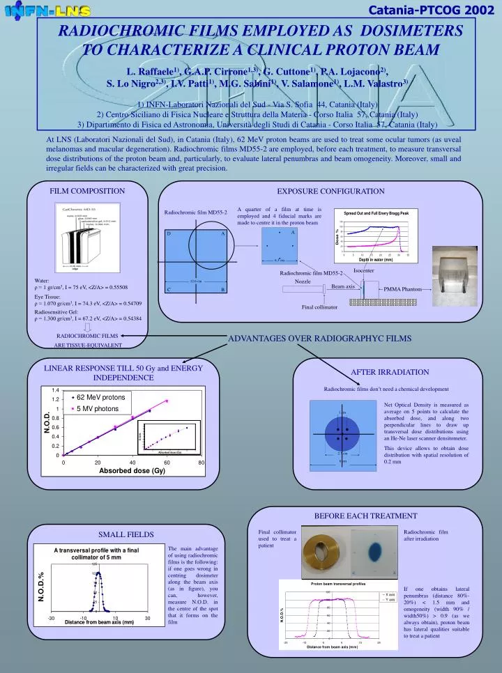
Catania-PTCOG 200 2. RADIOCHROMIC FILMS EMPLOYED AS DOSIMETERS TO CHARACTERIZE A CLINICAL PROTON BEAM L. Raffaele 1) , G.A.P. Cirrone 1,3) , G. Cuttone 1) , P.A. Lojacono 2) , S. Lo Nigro 2,3) , I.V. Patti 1) , M.G. Sabini 1) , V. Salamone 1) , L.M. Valastro 3)

E N D
Catania-PTCOG 2002 • RADIOCHROMIC FILMS EMPLOYED AS DOSIMETERS • TO CHARACTERIZE A CLINICAL PROTON BEAM • L. Raffaele1), G.A.P. Cirrone1,3), G. Cuttone1), P.A. Lojacono2), • S. Lo Nigro2,3), I.V. Patti1), M.G. Sabini1), V. Salamone1), L.M. Valastro3) • 1) INFN-Laboratori Nazionali del Sud - Via S. Sofia 44, Catania (Italy) • 2) Centro Siciliano di Fisica Nucleare e Struttura della Materia - Corso Italia 57, Catania (Italy) • 3) Dipartimento di Fisica ed Astronomia, Università degli Studi di Catania - Corso Italia 57, Catania (Italy) At LNS (Laboratori Nazionali del Sud), in Catania (Italy), 62 MeV proton beams are used to treat some ocular tumors (as uveal melanomas and macular degeneration).Radiochromic films MD55-2 are employed, before each treatment, to measure transversal dose distributions of the proton beam and, particularly, to evaluate lateral penumbras and beam omogeneity. Moreover, small and irregular fields can be characterized with great precision. FILM COMPOSITION EXPOSURE CONFIGURATION 6.3 cm A quarter of a film at time is employed and 4 fiducial marks are made to centre it in the proton beam Radiochromic film MD55-2 D A A Isocenter Radiochromic film MD55-2 12.6 cm Nozzle Beam axis C B PMMA Phantom Final collimator ADVANTAGES OVER RADIOGRAPHYC FILMS LINEAR RESPONSE TILL 50 Gy and ENERGY INDEPENDENCE RADIOCHROMIC FILMS ARE TISSUE-EQUIVALENT AFTER IRRADIATION Radiochromic films don’t need a chemical development 1 cm Net Optical Density is measured as average on 5 points to calculate the absorbed dose, and along two perpendicular lines to draw up transversal dose distributions using an He-Ne laser scanner densitometer. This device allows to obtain dose distribution with spatial resolution of 0.2 mm Water: ρ = 1 gr/cm3, I = 75 eV, <Z/A> = 0.55508 Eye Tissue: ρ = 1.070 gr/cm3, I = 74.3 eV, <Z/A> = 0.54709 Radiosensitive Gel: ρ = 1.300 gr/cm3, I = 67.2 eV, <Z/A> = 0.54384 2.5 cm 6 cm BEFORE EACH TREATMENT Final collimator used to treat a patient Radiochromic film after irradiation SMALL FIELDS The main advantage of using radiochromic films is the following: if one goes wrong in centring dosimeter along the beam axis (as in figure), you can, however, measure N.O.D. in the centre of the spot that it forms on the film If one obtains lateral penumbras (distance 80%-20%) < 1.5 mm and omogeneity (width 90% / width50%) > 0.9 (as we always obtain), proton beam has lateral qualities suitable to treat a patient