Download
1 / 1
10 likes | 169 Vues
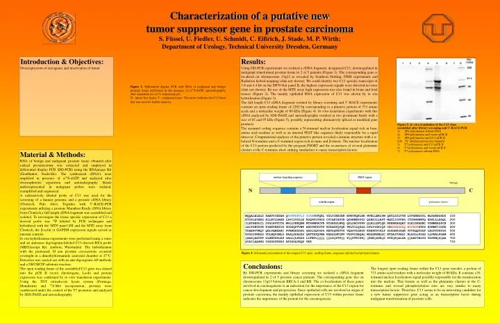
Figure 5: In vitro translation of the C13 clone assembled after library screening and 5’-RACE PCR . 1) SP6 polymerase wihout DNA 2) SP6 polymerase and vector pCR II 3) SP6 polymerase and C13-pCR II 4,8) 14 C labeled protein size standard

E N D
Figure 5: In vitro translation of the C13 clone assembled after library screening and 5’-RACE PCR. 1) SP6 polymerase wihout DNA 2) SP6 polymerase and vector pCR II 3) SP6 polymerase and C13-pCR II 4,8) 14C labeled protein size standard 5) T7 polymerase and C13-pCR II 6) T7 polymerase and vector pCR II 7) T7 polymerase wthout DNA nuclear targeting sequence PEST region 733 AA C N a-helix region glutamine cluster MQQALELALD RAEYVIESAR QRPPKRKYLS SGRKSVFQKL YDLYIEECEK EPEVKQKLRR NVNLLEKLVM QETLSCLVVN LYPGNEGYSL MLRGKNGSDS 100 ETIRLPYEEG ELLEYLDAEE LPPILVDLLE KSQVNIFHCG CVIAEIRDYR QSSNMKSPGY QSRHILLRPT MQTLICDVHS ITSDNHKWTQ EDKLLLESQL 200 ILATAEPLCL DPSIAVTCTA NRLLYNKQKM NTRPMKRCFK RYSRSSLNRQ QDLSHCPPPP QLRLLDFLQK RKERKAGQHY DLKISKAGNC VDMWKRSPCN 300 LAIPSEVDVE KYAKVEKSIK SDDSQPTVWP AHDVKDDYVF ECEAGTQYQK TKLTILQSLG DPLYYGKIQP CKADEESDSQ MSPSHSSTDD HSNWFIIGSK 400 TDAERVVNQY QELVQNEAKC PVKMSHSSSG SASLSQVSPG KETDQTETVS VQSSVLGKGV KHRPPPIKLP SSSGNSSSGN YFTPQQTSSF LKSPTPPPSS 500 KPSSIPRKSS VDLNQVSMLS PAALSPASSS QRTTATQVMA NSAGLNFINV VGSVCGAQAL MSGSNPMLGC NTGAITPAGI NLSGLLPSGG LLPNALPSAM 600 QAASQAGVPF GLKNTSSLRP LNLLQLPGGS LIFNTLQQQQQQLSQFTPQQ PQQPTTCSPQ QPGEQGSEQG STSQEQALSA QQAAVINLTG VGSFMQSQAA 700 AVAILAASNG YGSSSSTNSS ATSSSAYRQP VKK 733 Figure 4: Schematic presentation of the longest C13 open reading frame, sequence and derived protein features. Conclusions: By DD-PCR experiments and library screening we isolated a cDNA fragment downregulated in 2 of 5 prostate cancer patients. The corresponding gene lies on chromosome 13q13 between BRCA-2 and RB. The co-localization of these genes involved in carcinogenesis is an indication for the importance of the C13 region for cancer development and progression. Since epithelial cells are involved in origin of prostate carcinoma, the mainly epithelial expression of C13 within prostate tissue indicates the importance of the protein for the carcinogenesis. The longest open reading frame within the C13 gene encodes a protein of 733 amino acid residues with a molecular weight of 80 kDa. It contains a N-terminal nuclear localization signal possibly responsible for the translocation into the nucleus. This feature as well as the glutamine clusters at the C-terminus and several phosphorylation sites are very similar to many transcription factors. Therefore, C13 seems to be an interesting candidate for a new tumor suppressor gene acting as an transcription factor during malignant transformation of prostatic cells. Characterization of a putative new tumor suppressor gene in prostate carcinoma S. Füssel, U. Fiedler, U. Schmidt, C. Eißrich, J. Stade, M. P. Wirth; Department of Urology, Technical University Dresden, Germany Introduction & Objectives: Overexpression of oncogenes and inactivation of tumor . Results: Using DD-PCR experiments we isolated a cDNA fragment, designated C13, downregulated in malignant transformed prostate tissue in 2 of 5 patients (Figure 1). The corresponding gene is localized on chromosome 13q13 as revealed by Southern blotting, FISH experiments and Radiation hybrid mapping (data not shown). We could identify two C13 specific transcripts of 3.0 and 4.4 kb on the MTN blot panel II, the highest expression signals were detected in testes (data not shown). By use of the MTE array high expression was also found in brain and fetal tissues (Figure 2). The mainly epithelial RNA expression of C13 was shown by in situ hybridization (Figure 3). The full length C13 cDNA fragment isolated by library screening and 5’-RACE experiments contains an open reading frame of 2202 bp corresponding to a putative protein of 733 amino acids and a molecular weight of 80 kDa (Figure 4). In vitro translation experiments with this cDNA analyzed by SDS-PAGE and autoradiography resulted in two prominent bands with a size of 65 and 95 kDa (Figure 5), possibly representing alternatively spliced or modified gene products. The assumed coding sequence contains a N-terminal nuclear localization signal rich in basic amino acid residues as well as an internal PEST like sequence likely responsible for a rapid turnover. Computational analyses of the putative protein revealed a domain structure with a a-helical N-terminus and a C-terminal region rich in turns and b-sheets. The nuclear localization of the C13 protein predicted by the program PSORT and the occurrence of several glutamine clusters at the C-terminus show striking similarities to many transcription factors. Figure 1: Differential display PCR with RNA of malignant and benign prostate tissue performed in the presence of a35S-dATP, autoradiography after separation on a 6 % sequencing gel. N - tumor free tissue, T - malignant tissue. The arrow indicates the C13 band that was used for further analysis. Material & Methods: RNA of benign and malignant prostate tissue obtained after radical prostatectomy was extracted and employed in differential display PCR (DD-PCR) using the RNAimage Kit (GenHunter, Nashville). The synthesized cDNA’s were amplified in presence of a35S-dATP and analyzed after electrophoretic separation and autoradiography. Bands underrepresented in malignant probes were isolated, reamplified and sequenced. A radioactively labeled probe of C13 was used for the screening of a human genomic and a prostate cDNA library (Clontech, Palo Alto). Together with 5’-RACE-PCR experiments utilizing a prostate Marathon Ready cDNA library from Clontech a full length cDNA fragment was assembled and isolated. To investigate the tissue specific expression of C13 a derived probe was 32P labeled by PCR techniques and hybridized with the MTN panel I/II and the MTE array from Clontech, the b-actin or GAPDH expression signals served as internal controls. In situ hybridization experiments were performed using a sense and an antisense digoxigenin-labeled C13-derived RNA probe (MEGAscript Kit, Ambion, Wiesbaden). The hybridization with the pretreated 10 µm prostate cryosections occurred overnight in a dimethylformamide saturated chamber at 37°C. Detection was carried out with an anti-digoxgenin-AP antibody and a NBT/BCIP substrate reaction. The open reading frame of the assembled C13 gene was cloned into the pCR II vector (Invitrogene, Leek) and protein expression was confirmed by in vitro translation experiments. Using the TNT reticulocyte lysate system (Promega, Mannheim) and 35S-Met incorporation, proteins were synthesized under the control of the T7 promotor and analyzed by SDS-PAGE and autoradiography.