Download
1 / 78
780 likes | 881 Vues
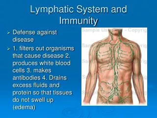
Lymphatic System and Immunity. Chapter 16. Functions of Lymphatic System. Draining interstitial fluid Transporting dietary lipids Protection. Lymphatic Vessels. Begin as closed ended lymph capillaries in tissue spaces between cells NOT A CIRCULATING FLUID

E N D
Lymphatic System and Immunity Chapter 16
Functions of Lymphatic System • Draining interstitial fluid • Transporting dietary lipids • Protection
Lymphatic Vessels • Begin as closed ended lymph capillaries in tissue spaces between cells • NOT A CIRCULATING FLUID • Interstitial fluid drains into lymphatic capillaries, forming lymph. • Lymph capillaries merge to form lymphatic vessels
Lymphatic vessels carry lymph into and out of lymph nodes • and finally back to the vascular system.
Lymphatic capillaries • Made of a single layer of squamous epithelial cells • Slightly larger than blood capillaries • Cells overlap and act as one-way valves • Opened by pressure of interstitial fluid • Anchoring filaments attach cells to surrounding tissue
Lymphatic vessels • Resemble veins (same 3 layers) • Found throughout body except: • Avascular tissues • Central nervous system • Splenic pulp • Bone marrow
Lymphatic vessels join to form lymphatic trunks. Lymphatic trunks join to form : Thoracic duct (3/4 of body) Right lymphatic duct (drains right arm, and right side of head, neck and upper torso) These empty into subclavian veins at junction with internal jugular vein.
Formation of lymph: Fluid leaves capillaries by diffusion and filtration Escaped proteins If lymph flow blocked = tissue swelling or edema Specialized lymphatic capillaries in vili of small intestine transport lipids - they are called lacteals, and the fluid is called chyle.
Lymphatic Organs • Red bone marrow Primary organs • Thymus gland • Lymph nodes • Lymph nodules Secondary organs • Spleen
Lymph Nodes • Lymph is filtered through lymph nodes • Found in clusters • “Waste water treatment plants” • Vary in size • Principal groupings in cervical, axillary and inguinal regions. • Provide biological filtration • Site of cancer growth and metastasis
Vessels enter node on convex side • Lymph passes through irregular channels called sinuses • Leaves node through one or two efferent vessels at the hilum or hilus • Capsule, cortex and medulla • Cortex contains lymph nodules • Follicular dendritic cells • Germinal centers – B cells proliferate
Lymph nodules are also found singly or in groups throughout the mucous membranes of the respiratory, urinary, reproductive and digestive tracts. MALT – mucosa associated lymphoid tissue Peyer’s patches in ileum Tonsils Some in appendix
Tonsils – lymphoid tissue under the mucous membranes of the throat palatine tonsils pharyngeal tonsil – adenoid lingual tonsils First line of defense Tonsillectomy
Thymus gland • in mediastinum above the heart • largest at age 10-12 then begins to atrophy • Pre - T cells come from bone marrow and develop into T cells • T cells then go to other lymphatic tissues • Thymus produces hormone thymosin - aids maturation of T cells elsewhere in body
Spleen • Largest lymphoid organ • In upper left quadrant of abdomen • Has a hilum and a capsule • Sinuses contain blood instead of lymph
White pulp: little islands, mostly B cells Red pulp: Venous sinuses Splenic cords – RBCs, macrophages, lymphocytes, plasma cells and granulocytes
Functions of Spleen • Blood formation – • All blood cells in fetus • Only lymphocytes and monocytes after birth • Blood filtration • Removes bacteria, particles, worn out RBCs and platelets (recycles iron) • Blood storage • Can contain over one pint of blood
Nonspecific Resistance The ability to ward off disease is called resistance. Lack of resistance is susceptibility. Nonspecific resistance refers to a wide variety of body responses against a wide range of pathogens. A pathogen is any microorganism that causes disease.
Immunity Immunity involves activation of specific lymphocytes to combat a specific foreign agent.
Nonspecific Resistance Species (Inborn) Resistance – certain species contract certain diseases, while other species do not.
Mechanical Barriers • Skin and mucous membranes : • First line of defense • Physical barrier • Shedding of dead cells • Mucus • Hairs • Cilia • Coughing and sneezing, production of tears, saliva, urine, defecation and vomiting physically remove harmful substances
Chemical Protection Sebaceous glands produce sebum – fatty acids inhibit growth of bacteria and fungi Lactic acid further decreases skin pH Accumulation of salt Vaginal secretions are also slightly acidic Gastric juice – acid, enzymes and mucus Lysozyme in tears, perspiration, saliva and tissue fluids
Normal Microbiota – bacteria living on skin inhibit the growth of pathogens by producing antibiotics
Antimicrobial Substances Transferrins are proteins that tie up the free iron in the blood and interstitial fluid. Interferon – “Paul Revere Chemical” – a glycoprotein produced by virus infected cells that cause neighboring cells to produce anti-viral proteins. These also enhance phagocytosis and can suppress growth of tumor cells.
The Complement System: • 10- 20 normally inactive proteins • When activated, they “complement” or enhance certain immune, allergic and inflammatory reactions. • Activation of inflammation • Opsonization – enhances phagocytosis • Cytolysis – membrane attack complex
Fever : Causes liver and spleen to sequester iron Increases phagocytosis Inhibits growth of microbes Speeds up body repair
Inflammation: Characterized by: Heat, swelling, redness, and pain (and sometimes loss of function) calor, tumor, rubor and dolor
Stages of inflammation • Vasodilation and increased permeability of blood vessels • Phagocyte migration • Neutrophils come first • Followed by macrophages • Tissue Repair
Phagocytosis Three phases: 1. Chemotaxis 2. Adherence 3. Ingestion
Natural Killer Cells • Next line of defense (with phagocytes) • Lymphocytes – but do not respond to specific antigens • Can kill a variety of microbes plus tumor cells. • May release perforins, or attack directly • Cell may not display correct MHC antigens
Immunity • Specific resistance to disease involving the production of a specific lymphocyte or antibody against a specific antigen. • An antigen is any substance that elicits an immune response. Best antigens are: • Large • Complex • Recognized as foreign
Haptens are molecules that are small, foreign and complex. To elicit an immune response, they must piggy-back on a larger molecule, often blood proteins. Epitopes: a foreign protein may result in several different antibodies. Each antibody recognizes a different portion of the protein. These regions are called epitopes.
Two forms of immunity: Humoral or antibody mediated immunity B cells (mature in bone) make antibodies: specific proteins that bind to specific antigens OR Cell-mediated immunity Tcytoxic lymphocytes attack virus infected or tumor cells directly