Download
1 / 76
760 likes | 934 Vues
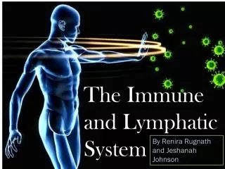
Lymphatic System and Immunity:. Lymphatic System. Lymph Lymphatic vessels Lymphatic tissue Lymphatic nodules Lymph nodes Tonsils Spleen Thymus. Lymphatic Vessels. Carry lymph away from tissues Lymphatic capillaries More permeable than blood capillaries

E N D
Lymphatic System • Lymph • Lymphatic vessels • Lymphatic tissue • Lymphatic nodules • Lymph nodes • Tonsils • Spleen • Thymus
Lymphatic Vessels • Carry lymph away from tissues • Lymphatic capillaries • More permeable than blood capillaries • Epithelium functions as series of one-way valves
Functions of the Lymphatic System • Fluid balance • Excess interstitial fluid enters lymphatic capillaries and becomes lymph • Fat absorption • Absorption of fat and other substances from digestive tract • Defense • Microorganisms and other foreign substances are filtered from lymph by lymph nodes and from blood by spleen
Lymphatic Tissue and Nodules • Lymphatic tissue • Consists mainly of lymphocytes • Encapsulated or not • Lymphatic nodules • Numerous in loose connective tissue of digestive (Peyer’s patches), respiratory, urinary, reproductive systems
Tonsils • Large groups of lymphatic nodules in nasopharynx and oral cavity • Provide protection against bacteria and other harmful material • Groups • Palatine • Pharyngeal • Lingual
Lymph Nodes • Organized in cortex and medulla • Substances removed by phagocytosis or stimulate lymphocytes or both • Only structures to filter lymph • Afferent and efferent vessels
Figure 25-16 Molecular Biology of the Cell (© Garland Science 2008)
Spleen • Located in left superior side of abdomen • Can be ruptured in traumatic abdominal injuries resulting in bleeding, shock, death • Blood flows through at 3 different rates • Fast (most), slow, intermediate • Functions • Destroys defective RBCs • Detects and responds to foreign substances • Limited reservoir for blood
Thymus • Located in superior mediastinum • Divisions: Cortex and medulla • Site of maturation of T cells
Hassall's corpuscle • Found in the central region of each thymic lobule • Sometimes referred to as a thymic corpuscle • Function is unknown • Known source ofThymic Stromal Lymphopoietin(TSLP) • TSLP is a Cytokine which activates Antigen Presenting Cells (APCs), which in turn play a strong role in T-lymphocyte selection.
Immunity • Ability to resist damage from foreign substances as microorganisms and harmful chemicals • Categories • Innate or nonspecific resistance • Mechanical mechanisms: Prevent entry or remove microbes • Chemical mediators: Promote phagocytosis and inflammation • Cells: Involved in phagocytosis and production of chemicals • Adaptive or specific immunity • Specificity: Ability to recognize a particular substance • Memory: Ability to remember previous encounters with a particular substance and respond rapidly
Innate immune Responses • Innate immune responses provide the first line of defense • The response lack specificity. • An invading agent first encounters a phagocytic cell. • Phagocytes have receptor proteins such as the Toll-like receptors (TLRs). • Activation of such receptors play a role in promoting immunity , by initiating secretion of inflammatory mediators (such as cytokines).
Innate responses are typically accompanied by the concentration of defensive agents at the site of infection—inflammation. Another mechanism produces proteins called complement that bind to pathogens and initiate their lysis. Innate responses against viruses include natural killer (NK) cell to induce apoptosis in the infected cell.
Inflammatory Response • Tissue injury regardless of type can cause inflammation • Response initiated by chemical mediators that produce vasodilation, chemotactic attraction, increased vascular permeability • Types • Local: Symptoms are redness, heat, swelling, pain, loss of function • Systemic: Symptoms are increase in neutrophil numbers, fever and shock
C3a stimulates mast cells and basophils, which then secrete inflammatory mediators
White blood cells Most important cellular components of immune system Methods Chemotaxis Phagocytosis Neutrophils Phagocytic and first cells to enter infected tissue Macrophages Monocytes that leave blood, enter tissues Large phagocytic cells Basophils and mast cells Promote inflammation Eosinophils Reduce inflammation Natural killer cells Lyse tumor and virus-infected cells Innate Immunity: Cells
Antigenic Determinants • Antigenic determinants • Specific regions of a given antigen recognized by a lymphocyte • Antigenic receptors • Surface of lymphocyte that combines with antigenic determinant
Another innate antiviral response is initiated by virus-infected cells that produce interferon. • Interferon binds to the surface of non-infected cells making them resistant to infection. • A type of interferon may induce synthesis of miRNAs that target viral RNA genomes.
Adaptive Immune Responses • Adaptive (or acquired) immune responses require a lag period for an attack against a foreign agent. • This response is specific and occurs only in vertebrates. • There are two broad categories of adaptive immunity: • Humoral immunity • Cell-mediated immunity
Humoral immunity is carried out by antibodies, which are globular proteins of the immunoglobulin superfamily (IgSF). Cell-mediated immunity is carried out by cells. Both types of immunity are mediated by lymphocytes, which are leukocytes that circulate between the blood and lymphoid organs.
Humoral immunity is mediated by B lymphocytes, which differentiate into antibody-secreting plasma cells when activated. Cell-mediated immunity is carried out by T lymphocytes (or T cells), which recognize and kill infected cells when activated. B and T cells arise from hematopoietic stem cells.
Origin and Developmentof Lymphocytes • B and T cells • Originate in red bone marrow • Move to lymphatic tissue from processing sites and continually circulate • Clones are small groups of identical lymphocytes
Adaptive immunity involves the ability to recognize, respond to, and remember a particular substance (stimulant). • Stimulants • Antigens: Large molecules • Foreign: Not produced by body, introduced from outside • Self-antigens: Produced by body • Haptens: Small molecules and capable of combining • Types • Humoral or Antibody-mediated: B cells • Cell-mediated: T cells
Major Histocompatability Complex (MHC) • Most lymphocyte activation involves glycoproteins of cell surfaces called MHC molecules • Class I molecules display antigens on surface of nucleated cells, resulting in destruction of cells • Class II molecules display antigens on surface of antigen-presenting cells (APCs), resulting in activation of immune cells
The MHC proteins hold fragments of antigen in place on APCs. • The TCR interacts with an APC when it docks with MHC proteins. • Cytotoxic T cells recognize their antigen in association with MHC I molecules. • Helper T cells recognize their antigen in association with MHC II molecules.
Peptides produced by antigen processing bind within a groove of the MHC protein molecule
Origin and Developmentof Lymphocytes • Positive selection • Ensures survival of lymphocytes that react against antigens • Negative selection • Eliminates lymphocytes that react against self-antigens • Primary lymphatic organs (red bone marrow, thymus) • Where lymphocytes mature into functional cells • Secondary lymphatic organs • Where lymphocytes produce an immune response
Thymus • Located in superior mediastinum • Divisions: Cortex and medulla • Site of maturation of T cells
T-cells, activated by clonal selection, interact with antigens through a surface protein called a T-cell receptor. • T cells are activated by fragments of antigens that are displayed on the surface of antigen-presenting cells (APCs). • Dendritic cells ingest antigens by endocytosis. • Macrophages ingest antigens by phagocytosis. • These cells process and present the antigen to other cells.
T cells release cytokines that alter the activity of the target cell.
Three classes of T cells are distinguished by the proteins on their surfaces and their biological functions: • Cytotoxic T lymphocytes (CTLs) kill target cells by inducing apoptosis. • Helper T (TH) lymphocytes are regulatory cells activated by APCs. • Regulatory T lymphocytes (TReg cells) suppress the activities of other immune cells.
T-cell receptor synthesis The ability of T cells to recognize foreign antigens is mediated by the T-cell receptor (TCR). Unlike most genes, the TCR gene is made up of a series of alternative gene fragments. In order to create a functional T cell receptor, immature T-lymphocyte precursors use a series of DNA-interacting enzymes to bring separate gene fragments together. The outcome of this process is that the TCR for EACHandEVERY T-lymphocyte has a different sequence.
DNA rearrangements that lead to the formation of genes for an immunoglobulin (such as the T-cell receptor)
DNA Rearrangement of Genes Encoding B- and T-Cell Antigen Receptors • Two separate genes (a C gene and V gene) are combined (with a joining segment) through rearrangement to form one continuous gene that encodes one antibody chain.
DNA rearrangement (continued) • The process is catalyzed by V(D)J recombinase which joins V and J segments of the gene, and deleting the intervening DNA. • Rearrangement is facilitated by signal sequences which are similar in V and J segments.
DNA rearrangement (continued) • Variability in polypeptide chains is achieved by: • The variety of V and J exons in the DNA of the germ line. • Varying the site at which J and V sequences are joined. • The enzymatic insertion of nucleotides. • Somatic hypermutation refers to a high mutation rate in V elements of B cells.