Download
1 / 20
220 likes | 421 Vues
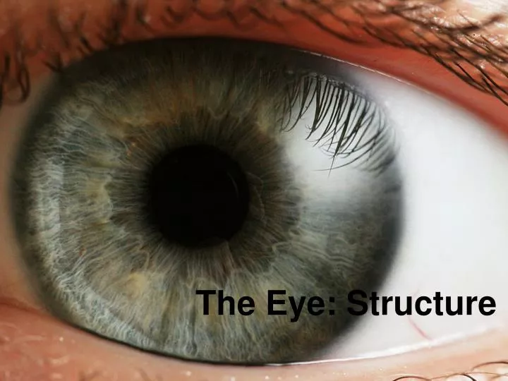
The Eye: Structure . 1. Sclera . White part of the eye Is made of tough tissue Protects the inner workings of the eye and gives the eye its shape. 2. Aqueous Humor. A clear, watery fluid Circulates in the front part of the eye, maintaining a constant pressure inside the eye. . 3. Cornea .

E N D
1. Sclera • White part of the eye • Is made of tough tissue • Protects the inner workings of the eye and gives the eye its shape
2. Aqueous Humor • A clear, watery fluid • Circulates in the front part of the eye, maintaining a constant pressure inside the eye.
3. Cornea • The curved, clear, outer surface • Covers the iris and pupil
4. Pupil • Is the opening in the center of the iris • The size of the pupil: • determines the amount of light entering the eye • controlled by circular muscles
5. Iris • The colored part of the eye • Made of circular muscles • Controls the size of the pupil
6. Lens • A clear convex lens • Is responsible for focusing light to form an image on the retina • Can change shape to focus on nearby and distant objects
7. Vitreous Humor • A clear, jelly-like substance • Fills the eye behind the lens • Holds the structures of the eye in place • Maintains the shape of the eye
8. Optic Nerve • A thick bundle made of millions of neurons • Carries visual information from the retina to the brain.
9. Retina • A thin, translucent, light-sensitive tissue made of specialized cells called rods and cones • Receives images formed by the lens
10. Optic Disc • The point where the optic nerve enters the retina • NOT sensitive to light • Known as the “blind spot”
11. Tapetum • A shiny layer that lines the area behind the retina of many animals • Helps make animal eyes visible in the dark • Allows them to see better at night
Corrective Lenses (Glasses & Contacts) • Near Sighted • Far Sighted
Color Blindness • Your eyes have special cells called rods and cones • Rods – Light sensitive • Cones – Color Sensitive
Color Blindness • There are three main kinds of color vision defects. Red-green color vision defects are the most common. This type occurs in men more than in women. The other major types are blue-yellow color vision defects and a complete absence of color vision.
Color Blindness • Most of the time, color blindness is genetic. There is no treatment, but most people adjust and the condition doesn't limit their activities.
LASIK • 1. Create a flap of corneal tissue • 2. Remodel the cornea using the laser • 3. Reposition the flap