Download
1 / 72
940 likes | 3.56k Vues
Antibody structure and function. Parham – Chapter 4. Outline. Antigens Antibody structure Antigen-antibody interactions Applications - immunoassays Generation of antibody diversity Isotype switching. Immunoglobulins – membrane-bound and soluble receptors. Epitopes.

E N D
Antibody structure and function Parham – Chapter 4 H. HogenEsch, 2009
Outline • Antigens • Antibody structure • Antigen-antibody interactions • Applications - immunoassays • Generation of antibody diversity • Isotype switching H. HogenEsch, 2009
Immunoglobulins – membrane-bound and soluble receptors H. HogenEsch, 2009
Epitopes • Epitope (antigenic determinant) is the part of an antigen to which an antibody binds. • Most antigens have multiple epitopes (multivalent) • Usually carbohydrate or peptide. Fig. 2.9 http://micro.magnet.fsu.edu/cells/viruses/influenzavirus.html H. HogenEsch, 2009
Epitopes recognized by antibodies are usually located at the antigen’s surface. Fig. 2.8 H. HogenEsch, 2009
Conformational vs. linear epitopes H. HogenEsch, 2009
Epitopes heat, acid Conformational epitopes - destroyed by denaturation Linear epitopes - unaffected by denaturation H. HogenEsch, 2009
Epitope recognition H. HogenEsch, 2009
Haptens Small molecules that are not immunogenic by themselves, but can bind immunoglobulins or TCRs. Haptens can induce an immune response when linked to a larger protein (carrier). H. HogenEsch, 2009
Hapten Parham Fig. 10.25 H. HogenEsch, 2009 Fig. 12.26
Hapten Parham Fig. 10.26 Fig. 12.27 H. HogenEsch, 2009
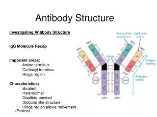
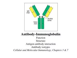
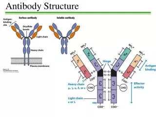
Basic structure of immunoglobulins • 2 light chains • lambda () • kappa (κ) 5 heavy chains - mu (μ) - gamma (γ) - alpha (α) - epsilon (ε) - delta (δ) Fig. 4.2 H. HogenEsch, 2009
Basic structure of immunoglobulins Fig. 4.2 H. HogenEsch, 2009
Antigen-binding Fragment Crystallizable Fragment H. HogenEsch, 2009
Hinge region provides flexibility to antigen-binding sites H. HogenEsch, 2009
Structure of immunoglobulins H. HogenEsch, 2009
Structure of immunoglobulins H. HogenEsch, 2009
Hypervariable and framework regions HV = CDR = complementarity -determining region Fig. 2.7 H. HogenEsch, 2009
Immunoglobulin classes (isotypes) L-chain: k or l H. HogenEsch, 2009
Differences between immunoglobulins H. HogenEsch, 2009
Allotypes • Small differences (few base pairs/amino acids) • May affect half life • May affect subclass distribution • Mendelian inheritance – autosomal dominant • Different distributions among ethnic groups • Associated with susceptibility to infectious diseases and autoimmune diseases H. HogenEsch, 2009
Antibody-antigen interaction H. HogenEsch, 2009 Fig. 2.10
Antibody-antigen interaction • Non-covalent binding: • Electrostatic • Hydrogen bonds • Van der Waals forces • Hydrophobic forces • Affinity: Strength of interaction between epitope and one antigen-binding site • Avidity: Strength of the sum of interactions between antibody and antigen Short range H. HogenEsch, 2009
Crossreactivity Antiserum raised against antigen A reacts also with antigen B Antigen A and B share epitopes Antigen A and B have similar (but not identical) epitopes H. HogenEsch, 2009
Crossreactivity H2N2 H2N3 Influenza virus H. HogenEsch, 2009
Monoclonal antibodies • Immortalization of a single clone of antibody-secreting cells • Fusion of B cells with neoplastic plasma (myeloma) cells H. HogenEsch, 2009
Monoclonal antibodies H. HogenEsch, 2009
Polyclonal vs. monoclonal antibodies • Polyclonal antibodies • purified from serum of immunized animals, often goats or rabbits. • Multiple specificities and affinities • Variation from batch to batch • Monoclonal antibodies • Produced by immortalized plasma cells, usually mouse origin. • Single specificity and affinity • Unlimited supply of identical antibody molecules H. HogenEsch, 2009
Examples of monoclonal antibodies as therapeutics H. HogenEsch, 2009
Types of therapeutic monoclonal antibodies H. HogenEsch, 2009
Rituximab in autoimmune disease(pemphigus vulgaris) Rituximab + IVGG Ahmed et al. NEJM, 355, 1772, 2006 Approximate cost: $3,976 per infusion or $15,904 for a four-dose course H. HogenEsch, 2009
Immunoassays • Precipitation assay • Agglutination assay • Enzyme-linked immunosorbent assay (ELISA) • Radioimmunoassay (RIA) • Western blotting • Immunofluorescence • Flow cytometry H. HogenEsch, 2009
Sensitivity of immunoassays precipitation - 30 mg/ml agglutination - 1 mg/ml radioimmunoassays, ELISA - 1 pg/ml H. HogenEsch, 2009
Precipitation reaction Aggregates formed by interaction of multivalent antibodies and multivalent macromolecular antigens. H. HogenEsch, 2009
Antigens have multiple epitopes H. HogenEsch, 2009
Hemagglutination H. HogenEsch, 2009
Coombs test • Direct: Add anti-human immunoglobulin antibodies (Coombs’ reagent) to red blood cells. Agglutination occurs if the red blood cells are coated with antibodies. • Indirect: Incubate test serum with red blood cells. Wash red blood cells. Add anti-human immunoglobulin antibodies. H. HogenEsch, 2009
Rhesus factor H. HogenEsch, 2009
1 2 3 4 Enzyme-linked immunosorbent assay (ELISA) Principle of ELISA/RIA • Coat wells with antigen • Add serum sample • Add enzyme-labeled • anti-human IgG • Add substrate H. HogenEsch, 2009
Western blotting Western blot H. HogenEsch, 2009
Immunofluorescence H. HogenEsch, 2009
Flow cytometry H. HogenEsch, 2009
Flow cytometry for CD4 T cells H. HogenEsch, 2009
Monitoring CD4 T cells in HIV infection H. HogenEsch, 2009
Immunoglobulin genes H. HogenEsch, 2009
V k J k C k germline DNA // 5’ 3’ 1 2 3 4 5 n 1 2 3 4 5 rearrangement 5’ 3’ B cell DNA V2J3 transcription 5’ 3’ primary RNA transcript splicing mRNA V2J3C translation k chain polypeptide V k C k V-region domains are constructed from gene segments H. HogenEsch, 2009
VL and VH-region domains are constructed from gene segments H. HogenEsch, 2009
Recombination Signal Sequences H. HogenEsch, 2009