Download
1 / 26
260 likes | 687 Vues
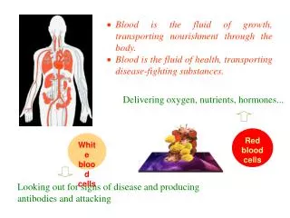
RED BLOOD CELLS. Red blood cells are one of the “formed elements of blood”. It is a circular, biconcave, non-nucleated disc. The edges are rounded and thicker than the centre, so when viewed from side it looks like a dumb bell. Erythropoiesis.

E N D
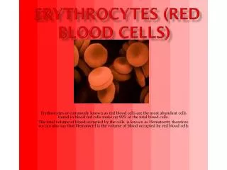
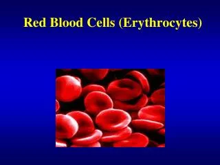
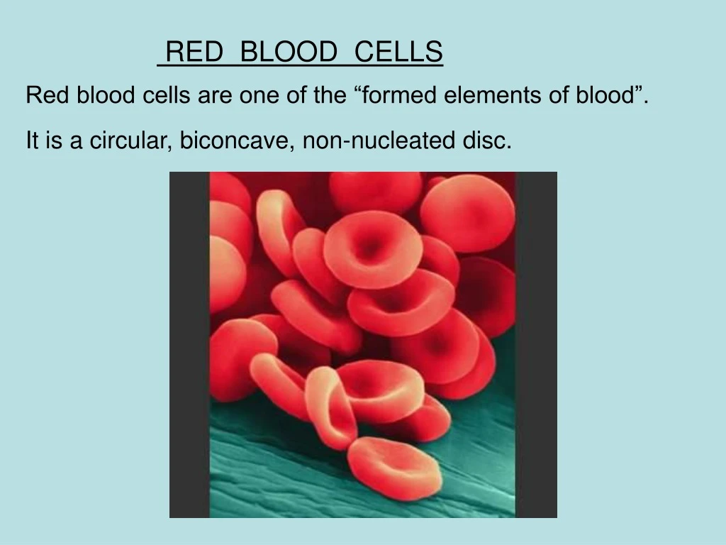
RED BLOOD CELLS Red blood cells are one of the “formed elements of blood”. It is a circular, biconcave, non-nucleated disc.
The edges are rounded and thicker than the centre, so when viewed from side it looks like a dumb bell.
Erythropoiesis Erythropoiesis is the process by which the origin, development and maturation of erythrocyte occur. Site of erythropoiesis: In fetal life:- 3 stages are seen. 1.Mesoblastic stage:- Up to 3 months of fetal life, RBCs are formed from mesoderm of yolk sac. In this stage erythropoiesis occurs in the blood vessels, so its also called intravascular erythropoiesis. 2. Hepatic stage:- From 3 months of fetal life, liver is the main site of blood formation 3. Myeloid stage:-During the last 3 mths of fetal life, liver and bone marrow are the site of formation.
In post natal life: In children– erythropoiesis occurs in All bones with red bone marrow (mainly) Liver and Spleen In adults—after 18 to 20yrs, from red bone marrow which includes: Ends of long bones Skull Vertebrae Ribs Sternum and Pelvis
Graph indicating the age and distribution of red bone marrow
Erythropoiesis in the bone marrow is called “medullary erythropoiesis” • Erythrtopoiesis in areas other than the bone marrow is called “extramedullary erythropoiesis” • Extramedullary erythropoiesis may occur in fetal erythropoietic tissue (liver and spleen) of an adult when the bone marrow cannot meet the physiologic needs of the tissues. This can lead to hepatomegaly and/or splenomegaly (increase in size of the liver or spleen because of increased functions in the organs).
Bone marrow – is located inside spongy bone • In a normal adult, ½ of the bone marrow is hematopoietically active (red marrow) and ½ is inactive, fatty marrow (yellow marrow). • The marrow contains both Erythroid (RBC) and leukocyte (WBC) precursors as well as platelet precursors. • Early in life most of the marrow is red marrow and it gradually decreases with age to the adult level of 50%. • In certain pathologic states the bone marrow can increase its activity to 5-10X its normal rate. • When this happens, the bone marrow is said to be hyperplastic because it replaces the yellow marrow with red marrow.
Hematopoiesis • All cellular elements derived from pluripotent stem cell (PPSC) • PPSC retains ability to both replicate itself and differentiate • About 75% of cells belong to WBC producing ‘myeloid series’ and only 25% belong to ‘erythroid series’ • Types of differentiation determined by the influence of various ‘Interleukins’ and ‘colony stimulating factors’.
Pluripoient stem cell Committed pluripotent stem cell Lymphoid stem cell Colony forming blastocyte Colony forming unit--M Colony forming unit--E Colony forming unit--GM megakaryocyte granulocyte M B E P N L E
Erythropoiesis Stages of erythropoiesis: • Proerythroblast • Early normoblast • Intermediate normoblast • Late normoblast • Reticulocyte • Mature RBC
Proerythroblast Cell size is about 22 microns, has both nucleoli and nucleus, no hemoglobin, on staining cytoplasm appears basophilic, confined to bone marrow and the cell can undergo mitotic cell division only under stress conditions.
Early normoblasts Cell size is about 16 microns, has only nucleus, no hemoglobin, on staining cytoplasm appears basophilic, confined to bone marrow and the cell demonstrates mitotic division
Intermediatenormoblast Cell size is about 14 microns, has nucleus, hemoglobin starts, on staining cytoplasm appears polychromatophilic, confined to bone marrow and the cell shows mitotic cell division.
Late normoblast Cell size is about 10 microns, nucleus becomes pyknotic then undergoes lysis and disappears, hemoglobin content increases, on staining cytoplasm appears eosinophilic, confined to bone marrow. From this stage onwards the cell division stops.
Is also known as immature RBC. Size is about 9 microns. Remnant of RNA appear as reticulum in the cytoplasm. No nucleus and hemoglobin content increases further. Staining property of cytoplasm is eosinophilic. The cells are present both in bone marrow and peripheral blood.
Mature erythrocyte Size is about 7.2 microns, lot of hemoglobin, cytoplasm staining is eosinophilic and present both in bone marrow and peripheral blood.
Some of the common features during erythropoiesis are • Gradual decrease in cell size. • Gradual disappearance of nucleoli and nucleus. • Arrest of mitotic division after the loss of nucleus. • Gradual increase in hemoglobin concentration. • Change in the staining property of cytoplasm.
The time required for mature RBCs to be formed from precursor stem cells is about 5-10 days and on an average it is about 7 days.
More about reticulocyte As already stated a reticulocyte is an immature RBC. It is about 20% larger than RBC. Is also a non nucleated cell. Retiulum is remnant of RNA. In adult the % of reticulocytes in circulation is about 0.5 to 1.0 and in newborn infant it is about 2 to 6. When the % of reticulocyte is more than normal, it is known as reticulocytosis.
What is the stimulus for erythropoiesis? • The most potent stimulus for erythropoiesis is hypoxia. In hypoxia oxygen supply to tissues is decreased. • Erythropoiesis is stimulated by the production of a substance called “erythropoietin” • Erythropoietin is a glycoprotein, secreted mainly by kidneys(85%) and also to certain extent by liver(15%). Its half life is about 5hrs
Formation and release of erythropoietin: Kidney Liver Hypoxia Renal erythropoietic factor Erythropoietinogen Erythropoietin Erythropoietin ( globulin) Erythropoietin stem cell Proerythroblasts
Mode of action of erythropoietin: • Causes early differentiation of proerythroblasts from pleuripotent stem cells in the bone marrow, and causes these cells to pass more rapidly through successive stages. Thus increases the RBC count in 2-3 days. • Increased synthesis of RNA, DNA, globins and ferritin which increases heam synthesis, thus increasing hemoglobin synthesis in already existing normoblasts. • Increases the release the reticulocytes from the bone marrow.
Factors necessary for erythropoiesis: • General factors— • Erythropoietin • Hormones-testosterone, thyroxin, growth hormone • and estrogen • Hemopoietic growth factor • Colony stimulating factor • Vitamins 2. Maturation factors— Vitamin B12, intrinsic factor and folic acid 3. Factors for Hb synthesis— Proteins and amino acids, iron, copper, cobalt and nickel ,vitamins.