Download
1 / 58
660 likes | 1.07k Vues
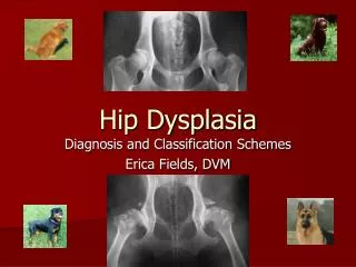
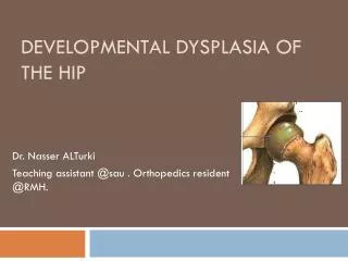
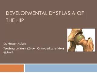
Developmental dysplasia of the hip (DDH). Definition Dysplasia of the hip that develop during fetal life or in infancy. It ranges from dysplasia of the acetabulum (shallow acetabulum) to subluxation of the joint to complete dislocation.

E N D
Definition • Dysplasia of the hip that develop during fetal life or in infancy. • It ranges from dysplasia of the acetabulum (shallow acetabulum) to subluxation of the joint to complete dislocation. • The old name was ‘‘congenital dysplasia of the hip (CDH).’’ The name has changed to indicate that not all cases are present at birth and that some cases can develop later on during infancy and childhood
Types: • DDH is classified into two major groups : • Typical and teratologic . • Typical DDH occurs in otherwise normal patients or those without defined syndromes or genetic conditions. • Teratologic hip dislocations usually have identifiable causes such as arthrogyposis or a genetic syndrome and occur before birth.
Developmental Dysplasia of the Hip 1. Complete hip dislocation. 2. Partial hip subluxation. 3. Hip dysplasia (incomplete development).
Incidence • Most newborn screening studies suggest that some degree of hip instability can be detected in 1/100 to 1/250 babies, actual dislocated or dislocatable hips are much less common, being found in 1-1.5 of 1000 live births. • There is marked geographic and racial variation in the incidence of DDH. • More inidence of DDH IN Sweden,Yugoslavia and Canada.
African and Asian caregivers have traditionally carried babies against their bodies in a shawl so that a child ’s hips are flexed, abducted, and free to move. • This keeps the hips in the optimal position for stability and for dynamic molding of the developing acetabulum by the cartilaginous femoral head. • Children in Native American and Eastern European cultures, which have a relatively high incidence of DDH, have historically been swaddled in confining clothes that bring their hips into extension. • This position increases the tension of the psoas muscle-tendon unit and might predispose the hips to displace and eventually dislocate laterally and superiorly.
Recommended Not recommended
Etiology • A positive family history for DDH is found in 12-33% of affected patients. • DDH is more common among female patients (80%). This is thought to be due to the greater susceptibility of female fetuses to maternal hormones such as relaxin, which increases ligamentous laxity • Primigravida. • Breech presentation(2-3%). • Oligohydramnios ,primigravida and large baby ( crowding phenomenon ). • Adduction and Extension postnatally.
Associated conditions • Torticollis • metatarsus adductus • calcaneovalgus feet
The left hip is the most commonly affected hip • In the most common fetal position, this is the hip that is usually forced into adduction against the mother’s sacrum. • Girls are affected 5 times more than boys.
CLINICAL FINDINGS • IN NEWBORNS • Usually asymptomatic and must be screened by special maneuvers • 1) Barlow test. It is a provocative test that attempts to dislocate an unstable hip. - Flexion ,adduction, posteriorly. - “Clunk”
Clinical Features : Neonates BARLOW’S TEST ( bahar lo)
Clinical Features : Neonates BARLOW’S TEST ( bahar lo)
2) Ortolani test It is a maneuver to reduce a recently dislocated hip. • Flexion, abduction, anteriorly. • We can`t use X-rays because the acetabulum and proximal femur are cartilaginous and wont be shown on X-ray. • US is the best method to Dx.
Clinical Features : Neonates ORTOLANI SIGN
Clinical Features : Neonates ORTOLANI SIGN
Clinical Manifestations • In infants: • As the baby enters the 2nd and 3rd months of life, the soft tissues begin to tighten and the Ortolani and Barlow tests are no longer reliable. • Shortening of the thigh, the Galeazzi sign , is best appreciated by placing both hips in 90 degrees of flexion and comparing the height of the knees, looking for asymmetry • Asymmetry of thigh and gluteal skin folds.
The most diagnostic sign is Ortolani’s limitation of abduction. • Abduction less than 60 degrees is almost diagnostic. • X-rays after the age of 3 months can be helpful esp. after the appearance of the ossific nucleus of the femoral head • US is 100% diagnostic.
Limitation of Abduction MOST RELIABLE SIGN
In walking child • In older children: Complaints of limping, waddling (bilateral DDH), lumbar lordosis, limitation of hip abduction, toe-walking, wide perineum, etc…
Screening • All neonates should have a clinical examination for hip instability • Risk factors : • breech presentation • family history • torticollis • oligohydramnios • metatarsus adductus USG SCREENING
CLINICAL & USG normal normal normal ABnormal ABnormal F/U till maturity REPEAT AT 6 WKS ABnormal normal Clinical & USG normal REPEAT AT 3 & 6 WKS ABnormal Closed / open reduction
DIAGNOSIS • 1. ULTRA SOUND • In the Graf technique, the transducer is placed over the greater trochanter, which allows visualization of the ilium, the bony acetabulum, the labrum, and the femoral epiphysis • The angle formed by the line of the ilium and a line tangential to the boney roof of the acetabulum is termed the α angle and represents the depth of the acetabulum. • Values > 60 degrees are considered normal, and those < 60 degrees imply acetabular dysplasia.
The β angle is formed by a line drawn tangential to the labrum and the line of the ilium; this represents the cartilaginous roof of the acetabulum. • A normal β angle is < 55 degrees, as the femoral head subluxates, the β angle increases.
Graf classification of DDH [ simplified]
X-ray • von rosen view: • hips abducted 45º &medially rotated. • Anteroposterior. • We draw a line through the central axis of the femoral shaft. in normal hip ( ossific nucleus )will be inside the acetabulum. in dislocated hip it will be above acetabulum.
X-ray • Horizontal line of Hilgenreiner: drawn between upper ends of tri-radiate cartilage of the acetabulum. • Vertical line of perkins: drawn from the lateral edge of the acetabulum vertical to horizontal line. • 4 quadrants: Normal hip: the ossification center of the femoral hip lower medial quadrant. Dislocated hip: upper lateral quadrant.
X-ray • Acetabular index: angle between horizontal line of hilgenreiner and the line between the two edges of the acetabulum. normal hip 20º30 dilocated or dysplastic hip ≥ 30º • Shenton’sline: semicircle between femoral neck and upper arm of obturator foramen, in dislocated hip this line is broken.
Treatment • The earlier the better. • Best time for treatment is in newborn period. • It depends on the device and age of the patient. • Goal is to: 1.Flex and abduct hips. 2.Reduce femoral head and maintaining it.
The goals in the management of DDH are to obtain and maintain a concentric reduction of the femoral head within the acetabulum to provide the optimal environment for the normal development of both the femoral head and acetabulum. • The later the diagnosisof DDH is made, the more difficult it is to achieve these goals, the less potential there is for acetabular and proximal femoral remodeling, and the more complex are the required treatments
Treatment • From (1-6 months) use Pavlik Harness. • From 6 months -2 year use hip spica. • From the age of >2 year traction , adductor tenotomy , surgical closed reduction, salter innominate osteotomy.
Infant 1 – 6 months of Age • First choice is PAVLIK harness • Ensure hip > 90 degrees flexion
Infant 1 – 6 months of age • weekly clinical examination & USG • By 3 weeks stable reduction must • Continue till radiographs show normal acetabulum Results : • 95% of initially dysplastic hips normal • 80% dislocated and not initially reducible were successfully reduced • Higher dislocations had a higher failure rate • The rate of AVN was 2.38%.
Pavlik harness • Standard of treatment worldwide • Upto 6 months • Contraindicated when there is major muscle imbalance (myelomeningocele,ligamentous laxity)
Complications of Pavlik Harness • AVN • Failure to reduce • Femoral nerve neuropathy • Inferior dislocation • Pavlik’s disease (flattening posterolateral acetabulum)
Child 6 months to 2 years of age • Closed or open reduction + adductor tenotomy • If closed reduction fails then surgeon should be prepared for an open procedure