Download
1 / 51
560 likes | 829 Vues
Hip Dysplasia and Developmental Dislocation of the Hip. dr n. med. Dariusz Mątewski. Definition. Incomplete developing of a hip joint within a prenatal period being able to be turn to dislocating

E N D
Hip Dysplasia andDevelopmental Dislocation of the Hip dr n. med. Dariusz Mątewski
Definition Incomplete developing of ahip joint within a prenatal period being able to be turn to dislocating Hip Dysplasia is a comprehensive term that has been used to include a spectrum of related developmental hip problems in infants and children, often present at birth.
Ethiology • Genetic factors • Ultraphysiological position of limbs of the foetus • Change of the foetus position • Laxity of joint capsule • Sudden extension of child’s limbs in hip joints Frequency range from 2 to 6 per 1000 new births
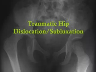
Classification: • Dysplasia without dislocation • Dysplasia with dislocation : • Subluxation • Dislocation
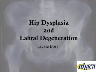
Pathology • structural changes in hip joint and surrounding tissues: • acetabulum oval, shallow, with increased antetorsion, „steep roof”, bold medial wall, filled by fatty and fibroblastic tissue • joint labrum reverse outside (subluxation and dislocation) • femoral head spherical shape, increased antetorsion , in the case of subluxation can be hyperthrophic (coxa magna) • joint capsulae hyperelastic, eliptic shape, elongation of ligamenum teres; • lack of hip joint stability: • Hyperelastic capsulae of hip joint • Abnormally development of acetabulum • subluxation or dislocation of femoral head.
Clinical tests • Ortolani’s test • Barlow’s test, • Asymetric abduction,
Clinical tests • Ortolani’s test • Barlow’s test, • Asymetric abduction,
Clinical tests • Ortolani’s test • Barlow’s test, • Asymetric abduction,
Clinical symptoms • Assymetry of hip rotations • Assymetry of gluteal plicaes • Positive Galeazzi symptom
Other symptoms of dislocation • Lack of femoral head in hip acetabulum; • Major trochanter shift above a Roser-Nelaton line (line between sciatic tuber and anterior superior illiac spine); • Shortening of a relative length of low lmb; • Limitation of abduction and external rotation • Positive Galeazzi’s test • Limping • Hyperlordosis • Static scoliosis
Other examinations • Sonographic examination (Graf method): • Static examination • Dynamic examination • X-ray examination (Hilgenreiner’s lines, Putti’s lines, Ombredanne`a – Perkins line, Calve’s line, Shenton-Menard’s line)
Goals of a conservative treatmentin newborns and infants • Extension of time, when hip joint is passed from flexion (foetus) to extension (newborn) • Preservation of joint congruency
Non-surgical: Wide position of thighs; Frejka’s pillow Pavlik’s harness; Van Rosen’s splint; Denis-Brown’s splint; Over head traction Surgical: Open reduction Pelvic procedures – pelvic osteotomy, acatabulum roof plasty, Femoral procedures –corective osteotomy decrease valgus position and antetorsion of femoral neck, Methods of treatment:
Conservative (non-surgical) treatment • Diagnosis immediately after birth: • Wide position of thighs, • Free position of limbs, • Pronation position of child in bed (after 6 week of life). • Diagnosis during 1 - 3 month of life: • Pavlik’s harness • Van Rosen’s splint • Frejka’s pillow • Koszla’s splint • Diagnosis after 6 month of life: • Denisa- Brown’s splint • Over head traction
Frog type plaster cast Over head traction
Treatment During first 6 months of life, dislocated hip joint can be reduction and treated by means of Pavlik’ harness. The most importance is proper using of the harness. Hip joint should be in flexion up to max. 110°, than abduction should be limited to intermediate position.
If after 2 – 3 weeks of using of Pavlik’s harness the hip joint is still dislocated, the using of this method of treatment should be cancelled. • If harness is function properly, it should be used by period equal to 2 times of age, when diagnosis od DDH was performed and additionaly 6 weeks. • If dysplasia was diagnosed in newborn immediately after birth, it should be treated by means of Pavlik harness by 6 weeks) .
90% of childrens with II° according to Graf criteria, which have diagnosed it immediately after birth – changes are being normalizing within 6 months without any kind of treatment. • Among children up to six month of life with positive Barlow test, treatment by means of Pavlik harness will be succesfull in 90%, and with positive Ortolani test will be succesfull in 80%.
Principles of DDH treatment by means of over head traction Skin traction (6 weeks) • Along the body axis (limbs are extented in the hip joints), • Along the axes of the limbs, which are flexed in hip joints under the right angle, • Gradual abduction of the lower limbs.
Principles of DDH treatment by means of over head traction • Plaster cast • Hip position in flexion and abduction (3 months) • Hip position in extension, abduction and internal rotation (Lange II) (next 2-3months)
Surgical treatment - indication • Symptoms of dysplasia, not recovered after conservative treatment, • Increased antetorsion of femoral neck, • Shallow acetabulum with „steep roof”,
Surgical procedures: -procedures on the pelvis: osteotomies: Salter, Pemberton, Dega,; -procedures on the femur: femoral derotational shortening;
Surgical technique –open reduction: -T shape capsular incision; -excision of the transverse ligamentum; - Removing of the ligamentum teres along with any associatedfatty tissue in the acetabularbase;
Surgical technique - acetabuloplasty: -Salter procedure, modification by Dega, or Pemberton; -redirects the acetabulum, thus immediately improving anterolateral coverage of the femoral head - Dega or Pemberton procedure - some risk of decreasingacetabular size because the techniqueincludes bending through the triradiatecartilage.
Surgical technique: - femoral derotational shortening osteotomy; - a goal: decreasing of pathological antetorsion ; - a incision leading from top of the major trochanter to the level of osteotomy; - using of pins determining the direction of osteotomy; - a wedge osteotomy;- osteosynthesis according to AO criteria
Complications • aseptic necrosis of the femoral head, • joint stiffness, • infection of surgical site. It is accepted, that if a reduction of hip joint dislocation is achievied before a first birthday, the risk of late complication is minimal. .
Goals of treatment of DDH: • reduction of dislocation; • restoring of a complete physical efficiency; • prevention of developing of degeneretive changes as sequele of dysplasia; • shortening of hospitalization time and minimalization of treatment costs;
a prognosis is good, if the treatment start early, • it will be worsening if the treatment delay or perform in an inappropriate way, as well as in the case of lasting of the disease. • prognosis will be poor and will lead to to the invalidity, if no treatment perform.