Download
1 / 1
10 likes | 138 Vues
Mouth. /e/. /u/. /a/. /i/. /o/. Notch. Chest - Accelerometer. Chest – Air coupled mic with BAI. Relationships between speech sounds and the subglottal airways: Preliminary Results. Graduate Student: Matías Zañartu

E N D
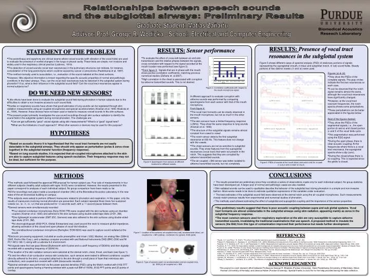
Mouth /e/ /u/ /a/ /i/ /o/ Notch Chest - Accelerometer Chest – Air coupled mic with BAI Relationships between speech sounds and the subglottal airways: Preliminary Results Graduate Student: Matías Zañartu Advisor: Prof. George R. Wodicka - School: Electrical and Computer Engineering Biomedical Acoustics Reseach Laboratory RESULTS: Presence of vocal tract resonances in the subglottal system RESULTS:Sensor performance STATEMENT OF THE PROBLEM • To evaluate the effect of cross-talk between air and skin transmission and the relative phases between the signals, cross-correlation with respect to the signal recorded at the mouth location was performed (see figure 3). • From figure 3: Signals that are in phase with the mouth show positive correlation coefficients, matching previous numerical studies (Zañartu et. al 2007). • High correlation in the chest is associated with corruption by airborne transmitted sounds. This is not desired. • The pectoriloquy and egophony are clinical exams where voiced sounds (with vibration of the vocal folds) are used to evaluate the presence of morbid changes in the lungs or pleural cavity. These tests are simple, non invasive and widely used in the respiratory clinical practice (Laënnec, 1819). • The detection of voiced sounds (vocal tract resonances) in the pulmonary structures can indicate, for instance, consolidation of the lung parenchyma which could be caused by cancer or pneumonia (Pasterkamp et al. 1997). • The method normally used is auscultation, i.e., evaluation of the sound radiated at the chest surfaces. • However, little objective information is known regarding the specific acoustic properties of normal and pathologic conditions in the lower airways. Thus, can the vocal tract resonances truly be detected in the subglottal system during phonation? If so, what is their influence in the subglottal sound field? Can the vocal tract resonances appear in normal subjects too? Figure 5 shows different cases of spectral analysis (PSD) of stationary portions of signals representing the supraglottal (mouth, in blue) and subglottal (notch, in red) sound fields. Steady portions of two distinct vowels (/i/ and /a/) were used. • Figures (a) & (d): • They show the PSDs of the complete signals. The gray circles indicate the first two resonances on each case. • It can be observed that the notch signal remains almost the same, although the vocal tract resonances have significantly changed. • However, at the vocal tract resonant frequencies, the notch spectra shows some perturbations. • These perturbations can be better appreciated in the figures below: • Rest of the figures (below): • They show the PSDs of the segmented signals, i.e., the open portion (b and e) and closed portion (c and f) of the vocal folds cycle. • This segmentation was performed using the EGG signal. • During the open phase, there is clear acoustic coupling: At the frequencies where there is a vocal tract resonance, the supraglottal spectra show a dip (zero). • During the closed phase there is no coupling. This is expected since the glottis is closed. Figure 3. Correlation coefficients with respect to the mouth microphone signal DO WE NEED NEW SENSORS? • A different approach to evaluate corruption with airborne sounds was performed by comparing spectrograms from each sensor with that of the mouth microphone. • From figure 4: • The vocal tract formants can be clearly observed in the mouth microphone, but not so much in the other sensors. • The skin sensors have a limited frequency response (<2kHz). They show the same response to lung sounds (Kraman et al. 1995) • The structure of the subglottal signals remains almost constant from vowel to vowel • The notch sensor detects the first subglottal resonance at 600 Hz. This feature does not change with the vowels. • The chest sensors are not as sensitive to subglottal sounds. They show features from the supraglottal resonances (vocal tract) that were not present in the notch. This suggests that they are corrupted by airborne transmitted sounds. • The air-coupled + BAI sensor was better isolated to airborne transmitted sounds, but not completely. • Little efforts have been done to evaluate the subglottal sound field during phonation in human subjects due to the difficulties to obtain a non invasive access to such sound field. • Studies on respiratory sounds have shown that good estimates of lung sounds can be registered through skin radiation measurements using air-coupled microphones and special accelerometers (Kraman et al. 1995; Wodicka et al., 1994). However, these devices have not been used or tested to measure voiced sounds in the skin surfaces. • The present project primarily investigates the use sound recordings through skin surface radiation to identify the sound field in the subglottal system during normal phonation. The challenges are: • Can we get sufficiently “good” voiced signals using skin measurements? What is a “good” signal here? • What are the limitations of such approach? What other sensors or devices may be used for this purpose? HYPOTHESES • Based on acoustic theory it is hypothesized that the vocal tract formants are not easily detectable in the subglottal airways. They should only appear as perturbation (poles & zeros close together) in the subglottal transfer function (frequency response). • Based on lung sounds studies, it is expected that air-coupled microphones attached to the skin are able to capture subglottal features using speech excitation. Their frequency response may not be ideal, but sufficient for the purpose. Figure 4. Spectrogram from sensors at different locations for different vowels. Figure 5. PSDs of sounds at the mouth (blue) and notch (red) for a vowel /i/ (a,b,c) and a vowel /a/ (d,e,f). CONCLUSIONS METHODS • The results presented are preliminary since they constitute a series of observations made only for each individual subject. No group statistics have been developed yet. A larger pool of normal and pathologic cases are also needed. • Skin radiated sounds can be used to qualitative describe the behavior of the subglottal tract during phonation in a simple and non-invasive fashion. More works needs to be done to estimate qualitative expressions of the variables under evaluation. • The best estimates of the subglottal sound field were observed at the sternal notch using the air-coupled microphones. Such measurements closely match previous results from experimental and numerical studies of phonation. • The methods used allowed estimating the effect of subglottal and supraglottal coupling and the importance of the sensor properties: • The methods used followed the approved IRB proposal for human subject use. Four sets of measurements in four different subjects (healthy adult subjects with ages 18-65) were considered. However, the results presented in this paper correspond to analyses of each individual subject. No group comparison have been made so far. • All the recordings took place inside a soundproof chamber (IAC) at the Biomedical Acoustics Laboratory in the new state-of-the-art biomedical building on campus. • The subjects were asked to perform different maneuvers related to phonation and respiration. In this paper, only the results of maneuvers involving normal phonation are presented. Each subject repeated three times five sustained vowels (/a/, /e/, /i/, /o/, /u/) that are produced for ~2 seconds each, with a ~1 second pause between them. • Several sensors were simultaneously recorded: • Four electret condenser microphones (Sony ECM-77B) were coupled with the skin surfaces using air chamber couplers (Kraman et al. 1995) and adhered to the skin surfaces using double-sided tape disks (2181, 3M). • One lightweight accelerometer (EMT 25C, Siemens) was also adhered to the skin surfaces using double-sided tape disks (2181, 3M). • An electroglottograph (EG2-PCX, Glottal Enterprises) was used around the neck to measure glottal impedance, allowing estimation of the closed and open phases of vocal fold vibration. • An omnidirectional condenser microphone (Beringher, ECM 8000) was used to capture sound radiated at the mouth. • Electronic processing equipment, included an audio preamplifier and mixer (1202, Mackie), an analog filter (3384 & 3343, Krohn-Hite Corp.), and a desktop computer provided with and National Instruments DAQ (BNC-2090 and NI PC1-M10-16E-1) along with a Labview 8.2 environment. • All signals were first low-pass filtered (Butterworth with 8 poles and a cutoff frequency of 5000Hz) and then digitally recorded with a sampling frequency of 22050 Hz. • The location of the skin radiation sensors were selected at the sternal notch, chest, throat, and back (see figure 1). • To test the effect of air-conduction versus skin conduction, such sensors were tested in different conditions: coupled (directly adhered to the skin), uncoupled (attached to the skin through a small piece of foam that minimizes skin conduction), and coupled but covered with a BAI (bioacoustic insulator). • Spectral estimation was performed via the power spectral densities (PSD) using the Welch method with 8192 FFT points and spectrograms having a Hanning window with a peak-null BW of 150Hz, 8192 FFT points and 20 points of overlap. • The preliminary results suggest that there is poor acoustic coupling between supra and sub glottal systems. Vocal tract formants are almost undetectable in the subglottal airways using skin radiation, appearing mainly as zeros in the subglottal frequency response. • The most common sensors used for respiratory exploration at the skin are very susceptible to capture airborne transmitted sounds, invalidating the traditional examinations that use speech. A proposed method to insulate the sensors (the BAI) from this type of contamination improved their performance but needs further development. Figure 1. Location of the sensors: air-coupled mics (red), accelerometer (blue), air-coupled mic + BAI (yellow), condenser mic (green), EGG (pink) REFERENCES • Austin, S. F. and Titze, I. R. (1997). "The effect of subglottal resonance upon vocal fold vibration," J. Voice 11, pp 391–402. • Harper, P., Pasterkamp, H., Kiyokawa, H., and Wodicka, G.R., (2003) “Modeling and measurement of flow effects on tracheal sounds,” IEEE Transactions on Biomedical Engineering, 50(1): 1-11. • Kraman, S. S., Wodicka, G. R., Oh, Y., and Pasterkamp, H. (1995): “Measurement of respiratory acoustic signals: Effect of microphone air cavity width, shape and venting”, Chest,108, pp. 1004–1008. • Laënnec, R. T. H. (1819) “De l'auscultation médiate ou traité du diagnostic de maladies des poumons et du coeur, fondé principalement sur ce nouveau moyen d'exploration“, Brosson et Chaudé, Paris. • Lulich, S. M., (2006) “The Role of Lower Airway Resonances in Defining Vowel Feature Contrasts”, PhD dissertation in Speech and Hearing Bioscience and Technology, MIT. • Pasterkamp, H., Kraman, S.S. and Wodicka, G.R., (1997) “Respiratory Sounds: Advances Beyond the Stethoscope“, Am J Respir Crit Care Med, Vol. 156. pp. 974–987. • Stevens, K. (2000). Acoustic Phonetics (MIT Press, Massachusetts), 1st ed. • Titze, I. R. and Story, B. H., (1997) “Acoustic interactions of the voice source with the lower vocal tract,” J. Acoust. Soc. Am, vol. 101(a), pp. 2234-2243. • Wodicka, G. R., Kraman, S. S., Zenk, G. M., and Pasterkamp, H. (1994): “Measurement of respiratory acoustic signals. Effect of microphone air cavity depth”Chest,106, pp. 1140–1144 • Zañartu, M., Mongeau, L. and Wodicka, G. R. (2007), "Influence of acoustic loading on an effective single mass model of the vocal folds", J. Acoust. Soc. Am., 121(2), pp. 1119-1129. • Zhang, Z., Neubauer, J. , and Berry, D. A. (2006), “The influence of subglottal acoustics on laboratory models of phonation”, J. Acoust. Soc. Am. 120(3), pp. 1558-1569. Figure 2. Type of sensors used: (a) accelerometers and air-coupled mics, (b) EGG,(c) air-coupled mic + BAI ACKNOWLEDGMENTS This project is a joint research effort between Professors George R. Wodicka (Purdue University), Hans Pasterkamp (University of Manitoba), Steve Kraman (University of Kentuky) and Jessica Huber (Purdue University). Special thanks to Julio Ho for the help provided during the data collection.