Download
1 / 49
500 likes | 516 Vues
FOETAL CIRCULATION. Philip Hassell , ST4. Characteristics of foetal circulatory dynamics. Parallel arrangement of two main arterial systems and their respective ventricles High impedance and low flow of pulmonary circulation Low impedance and high flow of placental circulation

E N D
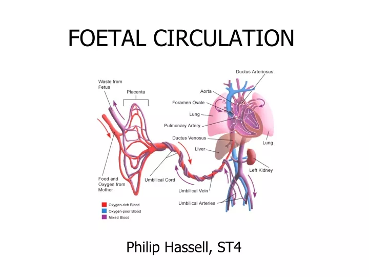
FOETAL CIRCULATION Philip Hassell, ST4
Characteristics of foetal circulatory dynamics Parallel arrangement of two main arterial systems and their respective ventricles High impedance and low flow of pulmonary circulation Low impedance and high flow of placental circulation Presence of shunts
Cardiac output and its distributionFoetal lamb CVO is 450 ml/kg/min UV flow is 200 ml/kg/min [45% of CVO] Of this,110 ml/kg/min [24%] passes through DV and 90 ml/kg/min [21%] passes through hepatic circulation
Cardiac output and its distributionFoetal lamb Total venous return to heart from IVC is 315ml/kg/min and represents 70% of CVO Of this 115 ml/kg/min [25% of CVO] passes through FO and 200 ml/kg/min [44%] passes through TV
Cardiac output and its distributionFoetal lamb Venous return to heart from SVC is 90 ml/mt/ and represents 21% of CVO most of this passes through tricuspid valve. RV ejects about 300 ml/mt or about 66% of CVO. About 35 ml/mt [8% of CVO] enters the pulmonary circulation
Cardiac output and its distributionFoetal lamb About 265 ml/kg/min [60%] passes through ductus arteriosus LV ejects 150 ml/kg [33%] Of this,90 ml/kg/min [20%] distributed to head and upper half and 45 ml/kg/min [10%] passes through isthmus 3% of CVO enters coronary circulation
60% 20% AA 33% 21% 8% 28 19 18 66% 70% 24% 24 70% 21% 32 45% 55%
Cardiac output and its distributionHuman foetus Limited data only is available based on doppler studies Umbilical blood flow is 180 ml/kg/min of estimated foetal weight Pulmonary blood flow is estimated to be 75ml/kg of foetal weight
Cardiac output and its distributionHuman foetus CVO appears to be similar to that in lamb, 450 ml/kg/min foetal weight
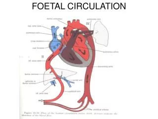
Venous return to heart Umbilical vein gives branches to left lobe of liver and then divides into DV and arcuate vein Arcuate vein joins the portal vein and then gives off branches to right lobe of liver Left hepatic vein joins the DV at the entry to IVC and right hepatic vein joins the IVC directly
Venous return to heart Posterior and left stream of IVC blood carries oxygenated blood while anterior and right stream carries poorly oxygenated blood Preferential streaming of DV and LHV blood across the foramen ovale and abdominal IVC and RHV blood across the TV
Venous return to heart Eustechian valve helps to direct the IVC blood to cross the foramen ovale The lower margin of septum secundum [christadividens] helps to direct the left posterior stream to preferentially cross the foramen ovale SVC blood is directed across the TV
Shunts in foetal circulation Ductus venosus Foramen ovale Ductus arteriosus or aortic isthmus
Shunts in foetal circulation The blood returning to heart through vena cavae is redistributed to tissues without being delivered to placenta represents effective R to L shunt The blood which passes through DV and then reaches DA and goes to placenta without getting distributed to tissues represent effective L to R shunt Combined R to L and L to R shunts forms 33% of CVO
Pulmonary Circulation Foetal lung does not serve gas exchange function PVR is high and PBF is low This helps to reduce workload of foetal heart
Pulmonary Circulation PA pressure rises gradually paralleling the rise in aortic pressure TPR falls gradually but this falls when correlated with rise in lung weight, there is actually an increase in PVR towards term PBF increases gradually
Pulmonary Circulation Experiments show foetal PBF increases dramatically in response to increase in maternal PaO2 This response is evident only in latter part of gestation
Pulmonary Circulation Breathing at birth is associated with a marked fall in PVR and rise in PBF PA pressure does not fall as rapidly and remains elevated until the DA has closed Once the ductus is closed, PA pressure can change independently of systemic pressure
Oxygen exchange function Higher haemoglobin level in foetus as compared to mother facilitates oxygen uptake by the foetus in the placenta Oxygen dissociation curve of foetal red cells is shifted to left as compared to adult red cells HbF has less affinity towards organic phosphates eg. 2,3-DPG
Oxygen exchange function These phosphates that are present in red cells compete with oxygen for binding to haemoglobin Affinity of reduced haemoglobin to 2,3-DPG is higher than that of oxyhaemoglobin and this facilitates oxygen delivery at tissue site
Oxygen exchange function As CO2 crosses the placenta from foetus to mother, it creates a local acidosis In the face of decreasing pH, mother’s haemoglobin shows less affinity towards Hb and oxygen release is enhanced [Bohr effect] This supports diffusion of more oxygen across the membrane to the foetus
Oxygen exchange function As O2 is released, maternal Hb acts as a buffer that removes H+ from local environment This encourages production of bicarbonate from H2O and CO2 thereby reducing local PaCO2 and facilitating diffusion of CO2 from foetus
Post-natal changes Gas exchange function is transferred from placenta to the lungs Separation of systemic and pulmonary circulations Increased metabolism to maintain body temperature and hence increased cardiac output
Post natal changes in various circulatory beds Coronary Blood flow decreases dramatically as the oxygen content increases Cerebral circulation also behaves in a similar fashion
Post natal changes in various circulatory beds Skin blood flow is high in-utero as the vessels are dilated because the skin is exposed to warm amniotic fluid Cutaneous vasoconstriction occurs post-natally as evaporation from skin starts Cutaneous flow falls and the vascular resistance increases
Post natal changes in various circulatory beds Hepatic blood flow falls rapidly post-natally with reduction in umbilical venous return and then increases as the GI flow is re-established Hepatic blood flow progressively increases after birth and by day 7 after birth reaches a level of 250 ml/min/100g by which time there is no flow through ductus venosus
Changes in Cardiac output Oxygen consumption increases from 6-8 ml/kg/min pre-natally to 15-20 ml/kg/min post-natally CVO of foetal lamb is 450 ml/kg/min CO of neonatal lamb is 300 -425 ml/kg/min, so the CVO will be 600 -850 ml/kg/min So the increase is 1.5 to 2 times
Changes in Cardiac outputMechanisms Neonate has to increase the metabolism to increase the body temperature as it is exposed to external temperature Improved diastolic function due to removal of compression by maternal organs and uterus causes increased cardiac filling and hence the cardiac output
Post-natal changes Gas exchange function is transferred from placenta to the lungs Separation of systemic and pulmonary circulations Increased metabolism to maintain body temperature and hence increased cardiac output
Changes in Cardiac outputMechanisms Peri-natal but not post natal increase in thyroid hormones is the principal mechanism for increase in cardiac output Improvement in myocardial growth and maturation brought about by cortisol may also play important role
Changes in haemoglobin and tissue oxygen delivery Human new born has a high haemoglobin level (about 16g/dl) so that the oxygen carrying capacity and the total amount of oxygen transported to tissues is high Since the HbF levels are still high facilitation at tissue site is not as great as in adults Over the first 8-10 weeks after the birth, Hb concentration falls to 10-11 g/dl. This is accompanied by loss of HbF and almost 100% is HbA
Regulation of foetal circulation Arterial baroreceptors are functional in the foetus from early in gestation Chemoreceptors are active only in latter part of gestation
Foetal circulation in pathological conditions Development of a structural abnormality will modify the foetal circulation This will affect the development of other components and can lead to other defects The impact of a defect will depend on it’s severity and time of gestation at which it occurs
Foetal circulation in pathological conditions Many of the defects, though it modifies the circulation, will not significantly affect foetal perfusion and hence the growth and development This is because of the presence of shunts and mixing of blood Foetus tolerates obstructive lesions greatly Yet the foetal circulation is jeopardized by regurgitant lesions and myocardial disease
Septal defects They in general do not modify the foetal circulation significantly VSD may have a transient left to right shunt in systole In ASDs, due to close proximity of defect with TV, more than normal amount of SVC blood may enter the LA.
Septal defects In atrioventricular septal defects, the obligatory flow from LV to RA will result in decrease in LV output and an increase in RV output This will reduce the flow across the isthmus and can predispose to co-arctation It is the degree of severity of AV valve lesion and regurgitation which will determine the outcome
Aortic arch abnormalities Most of the alteration in the circulation are due to co-existing intra cardiac defects Common features are, reduced flow in to ascending aorta, increased flow in to the pulmonary trunk and greater proportion of CVO carried across ductus to descending aorta The decreased volume loading of LV may possibly interfere with its development
TOF and related disorders Does not appear to affect foetal circulation adversely The volume and direction of flow across the PA and ductus are dependent on the severity of obstruction Total flow through the ductus will be reduced considerably
TOF and related disorders This can markedly reduce the diameter of foetal ductus and also reduce the development of smooth muscle in it’s wall If blood flows from aorta to PA in foetal life, the orientation of ductus changes and it forms an acute inferior angle with aorta AA and the isthmus carries larger than normal amount of blood and they tend to be larger
Ebstein’s anomaly Severe TR can manifest as in-utero cardiac failure especially if foramen ovale is restrictive Marked enlargement of RA and atrialised RV can cause septal displacement and compromise LV output Functional pulmonary atresia can result and ductal flow may be reversed
Ebstein’s anomaly Marked enlargement of right atrium can cause pulmonary hypoplasia Severe TR alters the preferential drainage of vena-caval blood and causes complete mixing of blood in right atrium