Download
1 / 55
720 likes | 2.07k Vues
Shoulder Pain. Marzena Slater, M.D. PGY 2 Emory Family Medicine. Objectives. To provide a background of shoulder anatomy Provide an overview of shoulder pain evaluation Discuss provocative testing used in the evaluation of shoulder pain

E N D
Shoulder Pain Marzena Slater, M.D. PGY 2 Emory Family Medicine
Objectives • To provide a background of shoulder anatomy • Provide an overview of shoulder pain evaluation • Discuss provocative testing used in the evaluation of shoulder pain • Summarize key history and physical findings that will aid in diagnosing common shoulder problems • Discuss common shoulder pathologies, their treatment and when to refer
Shoulder Anatomy Reference 1
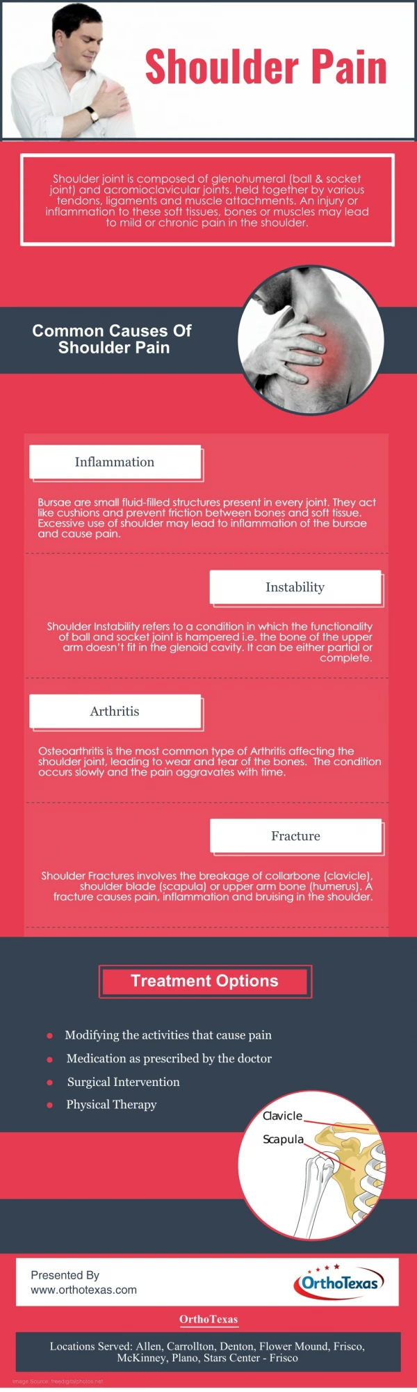
Shoulder Anatomy • The shoulder is one of the most complex joints in the body. • Composed of: 1.Bony structures: • Humerus • Glenoid • Acromion • Clavicle 2. Soft tissue structures • The rotator cuff muscles and supporting elements • 4 surrounding joints: • Glenohumeral joint • Acromioclavicular joint • Sternoclavicular joint • Scapulothoracic joint/pseudoarticulation
The Glenohumeral Joint • Most commonly dislocated major joint in the body • Basic principles: • GH joint a ball and socket joint • The glenoid fossa is flat and much smaller than the contacting humeral head (25-30%) • Cartilaginous labrum provides much of the socket function- but not much stability • Stability is achieved stabilizing structures
Static Stabilizers • Consist of: • Bony structures • Labrum • GH ligaments (superior, middle, inferior) • Joint capsule • Help maintain harmony • Continue to function in the presence of neurologic or intrinsic muscle pathology
Dynamic Stabilizers • Include • Rotator cuff • Scapular stabilizers (teres major, rhomboids, serratus anterior,trapezius and levator scapula) • Cannot function in the presence of neuromuscular injury or intrinsic muscle damage • Their malfunctioning leads to GH laxity and shoulder pain
Main function- depress the humeral head against the glenoid & stabilize Composed of 4 muscles: Supraspinatus- abduction helper to deltoid, pulls humeral head towards glenoid Infraspinatus- external rotation helper, pulls humeral head inferiorly Teres minor-external rotation helper, pulls humeral head inferiorly Subscapularis-internal rotation helper to pectoralis and latismus dorsi When damaged, humeral had can move upward within the joint 2/2 to unopposed deltoid action The Rotator Cuff
History • Always begin with the patient’s age, dominant hand and sport or work activity • Determine patient’s chief symptom (ex pain, weakness, instability, limited ROM) • How and when did the problem begin • Are patient’s sx related to specific injury/event or are the more insidious in onset • Do specific activities/arm movements exacerbate or alleviate the sx
History Cont. • Associated sx: • Instability/laxity (ex. Multidirectional GH instability) • Weakness (ex. Impingment, rotator cuff pathology) • Swelling (ex. Acute trauma/fx/rotator cuff tear) • Numbness (ex. C spine dz) • Loss of Motion/stiffness (ex. Adhesive capsulitis, dislocation or GH instability) • Catching (ex. Labral disorder) • Popping (ex. Labral disorder) • What previous treatments have been tried: ice, heat, medications (NSAIDS, Tylenol, ASA) • Previous medical interventions: physical therapy, injections, surgical interventions
Physical Exam • Approach it systematically • Don’t ignore the “good shoulder”- it can give you a reference for what’s normal in your patient • Expose both shoulders and perform: • Inspection • Palpation • ROM: passive and active in all planes • Strength testing- isolate relevant muscle groups individually • Special tests as indicated
Inspection • Look for: • Swelling • Asymmetry (ex. Squaring) • Muscle atrophy • Scars • Ecchymosis • Venous Distention
Inspection • Look for: • Swelling • Asymmetry (ex. Squaring) • Muscle atrophy • Scars • Ecchymosis • Venous Distention Ant. Shoulder Dislocation
Inspection • Look for: • Swelling • Asymmetry (ex. Squaring) • Muscle atrophy • Scars • Ecchymosis • Venous Distention Ant. Shoulder Dislocation AC joint separation
Inspection • Look for: • Swelling • Asymmetry (ex. Squaring) • Muscle atrophy • Scars • Ecchymosis • Venous Distention Ant. Shoulder Dislocation AC joint separation Supraspinatus and infraspinatus atrophy
Sternoclavicular joint Clavicle Coracoid process Acromion Acromioclavicular joint Scapula Palpation
Palpation • Bicipital Groove of biceps tendon • Subacromial Bursa • Cervical Spine
Range of Motion- Active • Apley “Scratch” Test is the quickest way to evaluate: • External rotation/ abduction (Fig 1) • Internal rotation/ adduction (Fig 2) • Internal rotation/ adduction (Fig 3) Fig 1 Fig 2 Fig 3
Range of Motion- Passive • If patient is unable to perform fully any of the active testing, passive testing should be conducted: • If Passive ROM normal but active ROM restricted, muscle weakness is a likely cause of restriction. • If passive and active ROM affected, bony (intra-articular) or soft tissue (extra-articular) blockage most likely- ex. Adhesive capsulitis
Range of Motion- Passive • Abduction- 180 degrees • Isolate the GH joint • 1st 20-30 degrees of abduction don’t require ST motion. • Arm internally rotated 1st 120 degrees (palm down) • Arm externally rotated (palm up) >120 degrees • Adduction- 45 degrees • Flexion- 90 degrees • Extension- 45 degrees • Internal Rotation- 55 deg • External Rotation- 40-45 deg.
Strength Testing- Rotator Cuff Evaluation • Always compare the 2 upper extremities • Isolate the rotator cuff muscle groups • Key finding with rotator cuff problems is pain accompanied by weakness. • True weakness should be distinguished from weakness due to pain
Supraspinatus • The “Empty can” test: • abduct shoulders to 90 degrees in forward flexion, with thumbs pointing down • The patient attempts to elevate arms against examiner resistance
Infraspinatus and Teres Minor • With patient’s arms at the sides, the patient flexes both elbows to 90 degrees while the examiner provides resistance against external rotation
Subscapularis • Lift off test: • Patient rests dorsum of the hand on the back in the lumbar area. • Inability to move hand off the back by further internal rotation of the arm, suggests injury to subscapularis muscle
Provocative Testing • Provide more focused evaluation for specific problems that you are suspecting from your initial H&P • Include: • Impingment signs: • Neer’s Sign, Hawkin’s Test • Rotator cuff tear • Drop Arm Test • AC joint Arthritis: • Cross-arm test • Cervical Nerve disorder: • Spurling’s Maneuver • GH instability: • Apprehension test, Relocation (Jobe), Sulcus Sign • Biceps Tendon instabillity/tendonitis: • Yergason test, Speed’s maneuver • Labral Disorders • Clunk Test, O’ Brien’s
Impingement Signs • Neer Sign • Arm in full flexion with arm fully pronated • Stabilize scapula • Pain= subacromial impingment-Rotator cuff tendons pinched under coracoacromial arch • Hawkins Test • Forward Flex shoulder to 90 deg., elbow@ 90 deg., then IR • Pain= suprapinatus tendon impingement or tendonitis • ? More sensitive for impingement than Neer’s Neer Neer Hawkins
Rotator Cuff Tear • Drop Arm Test: • Passively abduct patient’s shoulder to 90 degrees & have patient lower slowly to waist • Weakness or arm drop indicates rotator cuff tear/dysfunction • Note: the patient may be able to lower the arm slowly to 90 degrees (deltoid fxn) but will be unable to do so as far as the waist
AC joint pathology • Cross Arm Test: • Shoulder in 90 degrees forward flexion, then abduct arm across body • Pain indicates AC joint pathology • Decreased ROM indicates tight posterior capsule • AC Shear • Cup hands over clavicle/scapula: then squeeze • Pain/movement= AC pathology Cross Arm Test
Cervical Nerve Pathology • Pain that originates from the neck or radiates past elbow, is suspicious for neck disorder • Spurling Maneuver • Extend neck and rotate head of patient to affected shoulder. Then apply axial load. • Reproduction of sx indicates cervical disk pathology
GH instability • Apprehension test • Shoulder in neutral position at 90 deg abduction. • Apply slight ant pressure to humerus • Pain/apprehension about impending subluxation= GH instabiliy • Relocation (Jobe) • Perform if above positive • With patient supine, apply posterior force on prox. Humerus while externally rotating patient’s arm. • Decrease pain/apprehension= GH instability • Sulcus Sign • Arm to side, downward traction • Increased acromioclavicular sulcus= inferior instability Relocation Apprehension Sulcus
Biceps Tendonitis • Yergason’s • Patient’s elbow flexed at 90 deg with thumb up • Examiner grasps wrist, & resists patient attempt to supinate the arm and flex elbow • Pain= biceps tendonitis • Speed’s Maneuver • Flex pt’s elbow to 20-30 degrees w/ forearm in supination and arm in 60 degrees of flexion • Examiner resists forward flexion and palpates biceps tendon
Labral Disorders • Clunk Test • Patient supine • Patient’s arm is rotated & loaded from extension thru forward flexion. • “clunk sound” or clicking sensation, may indicate labral tear • OBrien’s • 90 deg FF, max IR, then adduct and flex
Clavicular Fractures • Common, most in mid 1/3 clavicle • Hx: • Fall on outstreched hand or direct blow • PE: • Point tenderness &/or a visible deformity • Always do neurovascular exam • Imaging • Xray- AP and cephalic tilt views • Rx: figure of 8 sling for 2-4 wks • F/U: in 4-6 wks with Xray • Refer to ortho: • If fx of distal clavicle- may disrupt ligaments of AC joint
Proximal Humeral Fractures • Hx: • Fall onto outstretched hand or direct blow • PE: • Crepitus at fx site • Ecchymosis within 24-48 hours of injury • Imaging: • AP and Lateral Xray. Axillary view if pt able • RX: • Shoulder immobilizer to prevent external rotation and abduction • Refer to ortho if: • Complex fx • Anatomic neck involvement • Displaced >1 cm • Ass. Neurovascular injury
Glenohumeral Dislocation • Most dislocations are anterior • Ant. Dislocation: • pt holds arm in external rotation/abduction • Humeral head palpable anteriorly/ dimple below acromion • Posterior Dislocation: • Arm in abduction/internal rotation • Dx often delayed • Imaging • Need two views: • AP- can miss posterior dislocation • Axillary or Y view AP Y view
Glenohumeral Dislocation • Complications: • Recurrent GH dislocations: • + apprehension, relocation or sulcus • Bony inury: • >50 % have Hill Sachs deformity- defect in posterolateral humeral head • Rotator Cuff Tear • 50% age <40, 80% >60 • RX: • Relocation • ROM exercises early • Recurrent- Bankart repair -surgical repair of detachement of labrum to glenoid
AC joint sprain/separation • Common injury among athletes and active patients • Mechanism: • direct blow to superior aspect of shoulder • lateral blow to deltoid area • Fall on outstretched hand • Exam: • Well localized swelling & tenderness over AC joint • Always examine pt in seated position • Palpable “stepped” deformity between the acromion and clavicle may indicate more severe injury • Imaging: • Xray: • AP- confirms dx • Axillary- if suspect grade 4-6 injury
Classification of AC injuries • Grade 3 and greater – refer to orthopedics for possible repair
Rotator Cuff Tear • Most common in greater than 40 yo. • Hx: • Younger patients- trauma related • Middle aged- chronic impingment leads to rupture of cuff • Imaging: • AP view GH joint- may show calcific tendonitis of cuff +/- superior migration of humeral head- should be f/u with further imaging • MRI= gold standard • RX: • Surgical repair in young and selected older patients within 3 weeks of injury preferably • Rehab for patients that are not surgical candidates
Impingement Syndrome • Mechanism: • rotator cuff tendons get impinged between coracoacromial arch and the humerus on abduction • Supraspinatus most commonly involved • Two types: • Primary • Older patient, chronic overuse and degeneration • Secondary • Younger, throwing athlete, GH instability leading to impingement
Impingement Syndrome • Hx: • Pain over anterolateral shoulder +/- radiation to elbow • Aggravated by overhead activities and worst at night • PE: +Hawkins, + Neer • RX: • Conservative: • Acute Phase: NSAIDS, Injection, Icing, rest • Pain resolving: Rotator cuff strengthening exercises • Xrays- get if 2-3 mo of conservative Rx fails- may show hooked acromion, AC spurring. • MRI as indicated • Surgery if conservative Rx fails
Frozen Shoulder • Mechanism: thickening and contracture of capsule around GH joint • Etiology: • Immobility (surgery, pain) • ?Autoimmune • Imaging: • X-rays- normal • Arthography- constriction of joint capsule • RX: • Physical therapy • Pain medications (NSAIDS) • Corticosteroids occasionally • Surgical referral if conservative fails The Origin of Acupuncture
Biceps Tendonitis • Inflammation of sheath around long head of biceps • Hx: • Pain and tenderness in bicipital groove • Often associated with impingement syndrome or rotator cuff tear • PE: +Yergason’s, +Speeds • Rx: • Conservative: Rest, ice, NSAIDs, Injection • Surgical: Transfer of tendon
Labral injury • SLAP lesion (Superior Labrum Anterior Posterior) common in throwing athletes • HX: Painful shoulder that clicks or pops with motion • PE: +clunk test, +O'Brien's, +/-laxity signs • Rx: • Often will need surgical repair, especially if athlete.
Osteolysis of Distal Clavicle • If atraumatic, most common in weight lifters • Begins as stress fx & bone remodeling cannot occur due to continual stress on joint • Hx: • Dull Pain over AC joint • worst in beginning of exercise period • Aggravated by abduction of shoulder • Dx: • Xrays- osteopenia and lucency of distal clavicle • RX: • D/C load-bearing activity • Surgical: Resection of distal clavicle
Case 1 • 42 yo Male comes to your office complaining of Rt shoulder pain. He does not remember any specific injury, but has been playing tennis a lot over the past 4 months and tells you that “opposing players no longer fear his serve”. It is difficult and painful for him to reach overhead and behind him. Even rolling onto his shoulder in bed is painful. • PE shows full ROM, but with discomfort at end ranges of Flexion, abduction and internal rotation. There is significant pain when you place the shoulder in position of 90 degrees of flexion and then internally rotate. There is also moderate weakness on abduction and external rotation. The rest of the MS exam is normal.
The most likely diagnosis is: • Acromioclavicular sprain • Rotator Cuff tear • Adhesive Capsulitis • Rotator Cuff impingement • Cervical Radiculopathy
The most likely diagnosis is: • Acromioclavicular sprain • Rotator Cuff tear • Adhesive Capsulitis • Rotator Cuff impingement • Cervical Radiculopathy
The best initial treatment is: • Corticosteroid injection • Arthroscopic subacromial decompression • Strengthening and ROM exercises • Elbow sling • Cervical collar