Download
1 / 22
220 likes | 335 Vues
Blood. Week 7 Dr. Walid Daoud A. Professor. Anemia ____________________________. Anemia is decrease in O2 carrying capacity of blood due to: 1- Decreased number of RBCs: . Less than 4.5 million/mm 3 in males. . Less than 3.9 million/mm 3 in females.

E N D
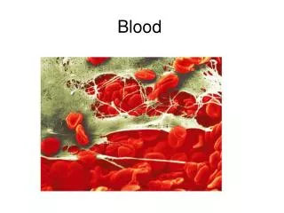
Blood Week 7 Dr. Walid Daoud A. Professor
Anemia____________________________ Anemia is decrease in O2 carrying capacity of blood due to: 1- Decreased number of RBCs: . Less than 4.5 million/mm3 in males. . Less than 3.9 million/mm3 in females. 2- Decreased Hb content of blood: . Less than 13.5 g/dl in males. . Less than 11.5 g/dl in females.
Classification & Causes of Anemia_____________________________ According to size of RBCs & Hb content: I-Normocytic Normochromic Anemia: Both size of RBCs & Hb content are normal but RBCs number is decreased. Causes: -Acute blood loss (Hemorrhagic anemia). -Bone marrow depression (Aplastic anemia). -Breakdown of RBCs(Hemolytic anemia).
Classification & Causes of Anemia____________________________ II-Microcytic Hypochromic Anemia or Iron Deficiency Anemia: Both size of RBCs and Hb contents are less than normal. Causes: -Decreased iron intake in diet. -Failure of iron absorption. -Chronic blood loss.
Classification & Causes of Anemia_____________________________ III-Macrocytic Hyperchromic Anemia or Megaloblastic Anemia: Both size of RBCs and Hb content are more than normal. Causes: -Vitamin B12 deficiency. -Folic acid deficiency.
Platelets or Thrombocytes____________________________ Non-nucleated, granulated bodies, 2-4 µm in diameter. Normal platelet count: 150,000-450,000/mm3 Decreased platelet count (Thrombocytopenia). Increased platelet count (Thrombocytosis).
Formation of Platelets____________________________ Pluripotential stem cell in bone marrow ↓ interleukin CFU-M ↓ GM-CSF Megakaryocyte ↓thrombopoietin Platelets CFU-M = Colony forming unit-megakayocyte. GM-CSF=Granulocye-monocyte colony stimulating factor.
Structure of Platelets___________________________ A. Platelet membrane. B. Platelet cytoplasm: 1- Beneath membrane: microtubules & ptn. 2- Intracellular organelles: 3- Glycogen granules. 4- Enzymes for synthesis of prostaglandins. 5- Granules: . Dense granules. . Alpha granules.
Hemostasis___________________________ It means stoppage of bleeding from an injured blood vessel. Hemostatic process consists of: I- Local vasoconstriction of injured blood vessel. II- Temporary hemostatic plug formation. III-Blood clot formation.
Hemostasis___________________________ I- Local vasoconstriction: 1- Nervous reflex. Initiated by pain sensation. 2- Local myogenic contraction. Due to direct damage of blood vessels. 3- Chemical substances. Serotonin and thromboxane A2 liberated from platelets cause vasoconstriction.
Hemostasis___________________________ II- Temporary Hemostatic Plug Formation: 1- Platelet adhesion. 2- Platelet activation. 3- Release reaction. 4- Platelet aggregation. 5- Platelet procoagulant activity. 6- Platelet fusion.
Hemostasis___________________________ III- Blood Coagulation (Blood Clotting): Properties of Blood Coagulation Factors:
Hemostasis___________________________ The clotting mechanism: The loose platelet plug changes to blood clot: 1-Fibrinogen forms a loose then tight fibrin clot catalyzed by factor XIII and Ca2+. 2-Thrombin converts fibrinogen to fibrin. 3-Active factor X converts prothrombin to thrombin. 4-Factor X is activated by either extrinsic or intrinsic pathways.
Hemostasis___________________________ A. Intrinsic Pathway: In vivo (in plasma) or in vitro (glass of tube). - Activation of factor XII by HMW kininogen and kallikrein in plasma. - XIIa activates factor XI. - XIa activates factor IX. - IXa forms a complex with VIIIa & activates factor X in presence of phospholipids (PF3) and Ca2+.
Hemostasis___________________________ B- Extrinsic Pathway: Initiated only in vivo by tissue thromboplastin (TPL, factor III); they are group of tissue phospholipoprotein released from damaged vascular and connective tissues. - TPL activates factor VII. - VIIa directly activates factor X in presence of Ca2+ TPL and phospholipids (PL) and indirectly through activation of factor IX.
Hemostasis___________________________ Common Part in Both Pathways: - Xa converts prothrombin to thrombin. - Thrombin transforms soluble fibrin to insoluble fibrin. - Thrombin in presence of Ca2+ activates factor XIII which stabilizes fibrin clot. - Contraction of actin, myosin and thrombosthenin of platelets causes clot retraction and squeezes serum out.
Hemostasis___________________________ Important notes: - Extrinsic pathway is very rapid (15 sec.) while intrinsic pathway is slow (1-6 min.). - Injury of a blood vessel will trigger both the intrinsic system by exposed collagen and extrinsic system by tissue thromboplastin. - In test tube, clotting occurs only by intrinsic system by glass or addition of collagen. - In intravenous thrombosis, clotting occurs via intrinsic system by exposure of clotting factors to subendothelial collagen.
Anticoagulant Mechanisms____________________________ Limiting reactions: prevent blood clotting in healthy blood vessels and breakdown any clots already formed. A.General:1-Smooth vascular endothelium. 2- Rapid flow of blood. 3- Heparin. B.Specific:1-Thromboxane A2 & Prostacyclin. 2- Antithrombin III. 3- Fibrinolytic system.
The Fibrinolytic System____________________________ Thrombomodulin binds thrombin to form thrombomodulin-thrombin system which activate protein C which causes: -Inactivation of factor Va and VIIIa. -Inactivation of inhibitor of tissue plasminogen activator (t-PA), ↑ formation of plasmin. Plasmin (fibrinolysin) is an enzyme formed from plasminogen. It lyses fibrin & fibrinogen forming fibrin-degradation products (FDP) which inhibits thrombin.
Anticoagulants____________________________ Substances used to prevent blood clotting. A- In vitro anticoagulants: in a test tube: 1-Removal of Ca2+ : . Oxalates: precipitates Ca2+ . . Citrates: binds Ca2+ by deionizing them 2-Silicon: prevents activation of factor XII. 3-Heparin.
Anticoagulants____________________________ B- In vivo anticoagulants: inside the body:
Abnormalities of Hemostasis____________________________ 1-Thrombocytopenic purpura: S.C. hemorrhage - ↓ Platelets count below 50,000 / mm3 . - Bleeding time is prolonged.. 2-Vitamin K deficiency: due to: -Decreased formation: absent intestinal flora. -Decreased absorption: bile duct obstruction. 3-Hemophilia: congenital sex-linked disease -Hemophilia A: absence factor VIII. -Hemophilia B: absence factor IX. -Hemophilia C: absence factor XI.