Download
1 / 1
10 likes | 120 Vues
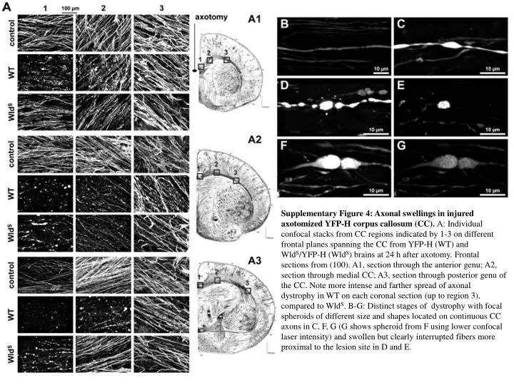
Supplementary Figure 4: Axonal swellings in injured axotomized YFP-H corpus callosum (CC). A: Individual confocal stacks from CC regions indicated by 1-3 on different frontal planes spanning the CC from YFP-H (WT) and

E N D
Supplementary Figure 4: Axonal swellings in injured axotomized YFP-H corpus callosum (CC). A: Individual confocal stacks from CC regions indicated by 1-3 on different frontal planes spanning the CC from YFP-H (WT) and WldS/YFP-H (WldS) brains at 24 h after axotomy. Frontal sections from (100). A1, section through the anterior genu; A2, section through medial CC; A3, section through posterior genu of the CC. Note more intense and farther spread of axonal dystrophy in WT on each coronal section (up to region 3), compared to WldS. B-G: Distinct stages of dystrophy with focal spheroids of different size and shapes located on continuous CC axons in C, F, G (G shows spheroid from F using lower confocal laser intensity) and swollen but clearly interrupted fibers more proximal to the lesion site in D and E.