Download
1 / 40
410 likes | 709 Vues
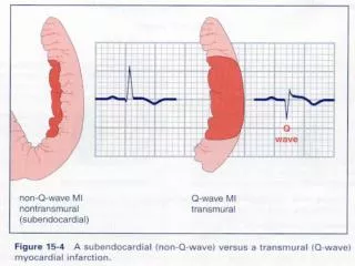
Vascular Disturbances III Infarction & Shock. Infarction. Infarction. Tissue necrosis due to ischaemia vascular insufficiency of any cause usually arterial occlusion due to thrombosis/embolism Mainly due to oxygen deficiency, but toxin accumulation & reperfusion injury may contribute

E N D
Infarction • Tissue necrosis due to ischaemia • vascular insufficiency of any cause • usually arterial occlusion due to thrombosis/embolism • Mainly due to oxygen deficiency, but toxin accumulation & reperfusion injury may contribute • Number of determining factors • Size of vessel and size of vascular territory • Partial / total vascular occlusion • Duration of ischaemia
Appearance of Infarct • Wedge-shaped • Occluded vessel at apex • Periphery of organ forms base • If extends to serosal surface, often overlying fibrinous exudate • Lateral margins blurred due to collateral blood supply
Appearance of Infarct NORMAL TISSUE ARTERY SURFACE FIBRINOUS EXUDATE INFARCTED TISSUE OCCLUSION ILL-DEFINED INFARCT BORDERS
Types of Infarct • Red (haemorrhagic) infarcts • Venous occlusion/congestion e.g. torsion • Loose tissues where haemorrhage can occur and blood can collect in infarcted zone e.g. lung • Tissues with dual blood supply e.g. lung small intestine (permitting blood flow from unobstructed vessel into infarcted zone – note flow is insufficient to rescue ischaemia) • Tissues that were previously congested due to sluggish venous outflow • When flow is re-established e.g. fragmentation of an occlusive embolus, angioplasty • White infarcts • arterial occlusion • solid tissues, where haemorrhage limited e.g. spleen, heart, kidney
Types of Infarct Red pulmonary infarcts - dual pulmonary / bronchial arterial supply
Types of Infarct White splenic infarct
Event Sequence • Coagulative necrosis • Infiltration by neutrophils • Infiltration by macrophages • Phagocytosis of debris • Granulation tissue formation • Scar formation
Event Sequence Day 1 3 7 14 90
Infarct, day 0 Fibre eosinophilia & contraction band necrosis
Infarct, day 1 Haemorrhage, necrosis and early neutrophil infiltrate
Infarct, day 3 Myocyte necrosis, pyknosis and marked neutrophil infiltrate
Infarct, day 7 Yellow necrotic infarct with hyperaemic border
Infarct, day 10 Granulation tissue after macrophage phagocytosis of infarcted cells
Infarct, day 90+ Subendocardial acellular fibrous scar
Infarct Development • Dependent on a number of factors • Nature of vascular supply • Dual supply e.g. lungs, liver • End arteries e.g. kidneys, spleen • Rate of vascular occlusion • Time for development of collateral circulation • Vulnerability to hypoxia • Neurons – 2-3mins, Myocardium – 20-30mins, Fibroblasts – hours. • Oxygen content of blood • Anaemia, cyanosis, congestive heart failure • Can result in infarction due to otherwise inconsequential blockage • Size of vessel and size of vascular territory • Partial / total vascular occlusion • Duration of ischaemia
Reperfusion Injury • Possible effects of re-establishing blood flow: • prevention of all necrosis • salvage of reversibly injured cells • accentuation of damage to irreversibly injured cells • new cellular damage • Latter two constitute reperfusion injury • Accentuated or new damage due to re-establishing blood flow • Many effects of ischaemic injury only seen when perfusion re-established
Reperfusion Injury • Can result in accelerated transition through stages of infarct development • Timing of infarcts unreliable post reperfusion • Inevitable if reperfusion occurs after optimum salvage time e.g. usually 6 hours after myocardial infarct • Characterised histologically by: • Marked haemorrhage • Marked contraction band necrosis
Reperfusion Injury • Causes: • delivery of oxygen and calcium ions to damaged tissue • interior of cells with damaged cell membranes exposed to high Ca++ conc cell lysis • generation of oxygen-dependent free radicals by damaged cells and phagocytes cell lysis • accentuation of O2-dependent damage • Anti-oxidants have only small effect on tissue loss • Acceleration of damage to irreversibly damaged cells more than new cellular damage
Shock (cardiovascular collapse) • Final Common Pathway for a umber of potentially lethal clinical events: • Severe Haemorrhage • Burns • Trauma • Large MI • (massive) Pulmonary Embolism • Microbial Sepsis
Shock (cardiovascular collapse) • Circulatory failure resulting in inadequate tissue perfusion (systemic hypoperfusion) • Results in: • hypotension • impaired tissue perfusion • cellular hypoxia • reversible cellular injury • irreversible cell injury and cell death
Types of Shock • Cardiogenic - due to myocardial pump failure • Intrinsic damage (MI) • Ventricular arrhythmias • Extrinsic compression (Tamponade) • Outflow obstruction (e.g. pulmonary embolism) • Hypovolaemic - due toloss of blood or plasma volume • Haemorrhage • Fluid Loss from severe burns • Trauma
Septic Systemic microbial infection Neurogenic Loss of vascular tone – spinal cord injury Anaphylactic Generalised IgE hypersensitivity response- systemic vasodilation - due to reduction in effective circulating blood volume peripheral pooling secondary to vasodilation and leakage of fluid due to increased vascular permeability
Types of Shock • Cardiogenic • myocardial pump failure • e.g. myocardial infarction, ventricular rupture, ventricular arrhythmia, cardiac tamponade, pulmonary embolism • Hypovolaemic • loss of blood or plasma volume • e.g. haemorrhage, trauma, burns, vomiting, diarrhoea
Types of Shock • Neurogenic shock • peripheral pooling of blood due to loss of vascular tone • e.g. anaesthetic accident / spinal cord injury • Anaphylactic shock • systemic IgE-mediated hypersensitivity reaction to allergens e.g. bee stings, peanut • release of mast cell mediators • systemic vasodilation and increased vascular permeability
Types of Shock • Septic shock • overwhelming microbial infection • gram negative sepsis • due to lipopolysaccharide (LPS / endotoxin) in walls • gram-positive / fungal septicaemia • due to molecules similar to LPS in walls • Super-antigen release
Septic Shock • Usually due to lipopolysaccharide (LPS/endotoxin) in walls of gram negative bacteria • LPS consists of fatty acid core and complex carbohydrate coat • Similar molecules in walls of gram positive bacteria or fungi • results in • endothelial damage • complement activation • activation of macrophages with cytokine release
Event Sequence • Low doses: • Local effectsof LPS & primary mediators (IL-1, TNF) • Complement activation by LPS • Monocyte/macrophage activation by LPS • binding to surface receptors • production of low doses of IL-1 and TNF • Endothelial cell activation by IL-1 & TNF • production of IL-6 & 8 by endothelium • increased adhesion molecule expression • Recruitment of inflammatory cells and cytokine cascade
Event Sequence • Intermediate doses: • Local effects of LPS & secondary mediators (NO, PAF) • Systemic effects of primary mediators (IL-1, TNF) • Endothelial cell injury by LPS • triggering of coagulation cascade • increased vascular permeability • production of secondary mediators by endothelium • Local vasodilation due to secondary mediators: nitric oxide, platelet activating factor • Systemic effects of IL-1 and TNF • Fever • Acute-phase reactant production (CRP, fibrinogen)
Event Sequence • High doses • Systemic effects of LPS, primary & secondary mediators • Widespread endothelial cell injury (LPS, cytokines) • Acute respiratory distress syndrome • Widespread activation of coagulation (LPS, cytokines) • Disseminated intravascular coagulation • Peripheral vasodilation, decreased cardiac contractility (NO) • Hypotension • Multiorgan failure due to hypoperfusion
Multiorgan Failure • Multiple organ damage due to ischaemia secondary to hypoperfusion • brain: ischaemic encephalopathy • heart: subendocardial infarcts • kidney: acute tubular necrosis • GIT: haemorrhagic enteropathy / ischaemia • liver: fatty change / centrilobular haemorrhagic necrosis • ARDS in lungs commonly present concurrently • Due to microvascular injury, not ischaemia
Stages of Shock • Nonprogressive phase • Reflex compensatory mechanisms maintain perfusion of vital organs • Tachycardia, peripheral vasoconstriction (pale cold clammy skin), renal conservation of fluid (anuria) • Progressive phase • Tissue hypoperfusion & metabolic imbalance • Development of acidosis • Due to anaerobic glycolysis and renal failure • Causes arteriolar dilatation and peripheral pooling of blood • Worsens hypotension and exacerbates tissue ischaemia • Irreversible phase • Irreversible cellular and tissue injury • No response even if haemodynamic defects corrected
Consequences of Shock Pink hyaline membranes lining alveolar spaces in ARDS
Consequences of Shock ATN – swollen, sloughed and flattened regenerating tubular epithelium, normal glomerulus Normal glomerulus and tubules
Consequences of Shock • Prognosis varies with cause and duration • If circulatory disturbance corrected during nonprogressive phase full recovery • If progress to irreversible phase high mortality • Hypovolaemic shock • > 80 – 90% survival in young healthy adults (10 - 20% mortality) • Cardiogenic shock due to MI / Septic shock • up to 75% mortality even with appropriate management
Summary • Infarction • Definition • Infarct Types • Timing of Infarcts • Reperfusion injury • Shock • Types of shock and aetiology • Stages of shock • Consequences