Download
1 / 26
270 likes | 419 Vues
Lymphoma. ALL. CLL. Lymphomas. MM. Neutrophils. AML. Myeloproliferative disorders. Myeloid progenitor. Eosinophils. Hematopoietic stem cell. Basophils. Monocytes. Platelets. Red cells. na ï ve. germinal center. B-lymphocytes. Plasma cells. Lymphoid progenitor. T-lymphocytes.

E N D
ALL CLL Lymphomas MM Neutrophils AML Myeloproliferative disorders Myeloid progenitor Eosinophils Hematopoietic stem cell Basophils Monocytes Platelets Red cells naïve germinal center B-lymphocytes Plasma cells Lymphoid progenitor T-lymphocytes
CLL MM ALL DLBCL, FL, HL B-cell development memory B-cell germinal center B-cell stem cell mature naive B-cell lymphoid progenitor progenitor-B pre-B immature B-cell plasma cell Bone marrow Lymphoid tissue
Lymphoma classification(2001 WHO) • B-cell neoplasms • precursor • mature • T-cell & NK-cell neoplasms • precursor • mature • Hodgkin lymphoma Non- Hodgkin Lymphomas
Mechanisms of lymphomagenesis • Genetic alterations • Infection • Antigen stimulation • Immunosuppression
Epidemiology of lymphomas • 5th most frequently diagnosed cancer in both sexes • males > females • incidence • NHL increasing • Hodgkin lymphoma stable
Risk factors for NHL • immunosuppression or immunodeficiency • connective tissue disease • family history of lymphoma • infectious agents • ionizing radiation
Clinical manifestations • Variable • severity: asymptomatic to extremely ill • time course: evolution over weeks, months, or years • Systemic manifestations • fever, night sweats, weight loss, anorexia, pruritis • Local manifestations • lymphadenopathy, splenomegaly most common • any tissue potentially can be infiltrated
Other complications of lymphoma • bone marrow failure (infiltration) • CNS infiltration • immune hemolysis or thrombocytopenia • compression of structures (eg spinal cord, ureters) • pleural/pericardial effusions, ascites
Diagnosis requires an adequate biopsy • Diagnosis should be biopsy-proven before treatment is initiated • Need enough tissue to assess cells and architecture • open bx vs core needle bx vs FNA
Stage I Stage II Stage III Stage IV Staging of lymphoma A: absence of B symptoms B: fever, night sweats, weight loss
Three common lymphomas • Follicular lymphoma • Diffuse large B-cell lymphoma • Hodgkin lymphoma
Relative frequencies of different lymphomas Non-Hodgkin Lymphomas Diffuse large B-cell Hodgkin lymphoma NHL Follicular Other NHL ~85% of NHL are B-lineage
Follicular lymphoma • most common type of “indolent” lymphoma • usually widespread at presentation • often asymptomatic • not curable (some exceptions) • associated with BCL-2 gene rearrangement [t(14;18)] • cell of origin: germinal center B-cell
defer treatment if asymptomatic (“watch-and-wait”) • several chemotherapy options if symptomatic • median survival: years • despite “indolent” label, morbidity and mortality can be considerable • transformation to aggressive lymphoma can occur
Diffuse large B-cell lymphoma • most common type of “aggressive” lymphoma • usually symptomatic • extranodal involvement is common • cell of origin: germinal center B-cell • treatment should be offered • curable in ~ 40%
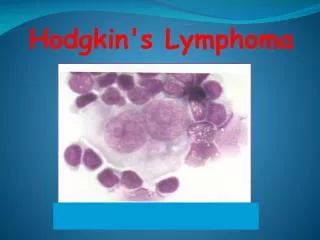
Hodgkin lymphoma • cell of origin: germinal centre B-cell • Reed-Sternberg cells (or RS variants) in the affected tissues • most cells in affected lymph node are polyclonal reactive lymphoid cells, not neoplastic cells
RS cell and variants classic RS cell lacunar cell popcorn cell (lymphocyte predominance) (mixed cellularity) (nodular sclerosis)
A possible model of pathogenesis loss of apoptosis transforming event(s) EBV? cytokines germinal centre B cell RS cell inflammatory response
Hodgkin lymphomaHistologic subtypes • Classical Hodgkin lymphoma • nodular sclerosis (most common subtype) • mixed cellularity • lymphocyte-rich • lymphocyte depleted
Epidemiology • less frequent than non-Hodgkin lymphoma • overall M>F • peak incidence in 3rd decade
Associated (etiological?) factors • EBV infection • smaller family size • higher socio-economic status • caucasian > non-caucasian • possible genetic predisposition • other: HIV? occupation? herbicides?
Clinical manifestations: • lymphadenopathy • contiguous spread • extranodal sites relatively uncommon except in advanced disease • “B” symptoms