Download
1 / 47
571 likes | 910 Vues
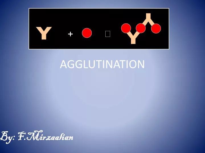
Y. +. . Y. Y. AGGLUTINATION. By: F.Mirzaalian. Tests Based on Ag/Ab Reactions. All tests based on Ag/Ab reactions will have to depend on lattice formation or they will have to utilize ways to detect small immune complexes

E N D
Y + Y Y AGGLUTINATION By: F.Mirzaalian
Tests Based on Ag/Ab Reactions • All tests based on Ag/Ab reactions will have to depend on lattice formation or they will have to utilize ways to detect small immune complexes • All tests based on Ag/Ab reactions can be used to detect either Ag or Ab
Complex may be directly visible or invisible Directly visible – agglutination Invisible • requires specific probes (enzyme-labelled anti-immunoglobulin, isotope-labelled anti-immunoglobulin, etc.) • binds Ag-Ab complex and amplifys signals • signals can be measured by naked eyes or specific equipment e.g. in ELISA, RIA, IFA
Methods for Ag-Ab detection • Precipitation • Agglutination • Hemagglutination and hemagglutination inhibition • Viral neutralization test • Radio-immunoassays • ELISA • Immunoflourescence • Immunoblotting • Immunochromatography
Agglutination 1- Active (direct) agglutination. 2- Passive (Indirect) agglutination. 3- Reverse Passive agglutination. 4- Hemagglutination 5- Hemagglutination inhibition. 6- Viral Hemagglutination. Precipitation(Immunodiffusion) A-Immunodiffusion in gel (Passive e.g RID- Active e.gelectroimmunodiffusion) B- Fluid phase Immunoprecipitation e.g. nephelometry and turbidimetry
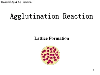
Lattice Formation Agglutination Tests
Agglutination • The interaction between antibody and a particulate(Insoluble) antigen results in visible clumping called agglutination. • Particulate antigen include: • bacteria, • white blood cells, • red blood cells, • latex particles
Agglutination Test positive. negative. antigen Antibody.
Phases of Agglutination • Agglutination is a two-Phase reaction that results in the formation of a stable lattice network • Primary Phase (Sensitization) • Ab reacts with a single antigenic determinant on the surface of Ag. • Secondary Phase (Lattice formation) • Ab must be able to bridge the gap between particles so that at least one Fab portion is attached to an antigenic determinant on each of two adjacent particles, is dependent on environmental conditions and the relative concentrations of antigen and antibody.
This represents what occurs during stage one of agglutination: Sensitization Stage 1 Antibody molecules attach to their corresponding Antigenic site (epitope) on the red blood cell membrane. There is no visible clumping.
This represents what occurs during stage 2 of agglutination: Lattice Formation Stage 2 Antibody molecules crosslink RBCs forming a lattice that results in visible clumping or agglutination.
Agglutinin and Agglutinogen • An agglutinin is an antibody that interacts with antigen on the surface of particles such as erythrocytes, bacteria, or latex particles to cause their agglutination. • An agglutinogen is an antigen on the surface of particles such as red blood cells that react with the antibody known as agglutinin to produce agglutination. • The most widely known agglutinogens are those of the ABO and related blood group systems.
The following examples of agglutination reactions : • 1. Rheumatoid factor latex agglutination • 2. Bacterial latex agglutination • 3. Coombs test • 4. Blood typing
Agglutination • In this test the antigen is particulate (visible, big and insoluble) (e.g. bacteria and red blood cells) or an inert particle (latex beads) coated with antigen. • Antibody is divalent and cross links the multivalent antigen to form a lattice network or clumps (agglutination). • This reaction can be performed in a tube or on a glass slide e.g. ABO blood grouping.
Slide agglutination is a rapid method to determine the presence of agglutinating abs
Factors influencing the reaction: • Elevation or decrease of temperature. • Motion (shaking,stirring,centrifugation). • PH. • Class of antibody (IgM/IgG).
When Abs & Ags are present in equimolar ratios They form insoluble complexes that ppt (ZONE OF EQUIVALENCE) Decreased amounts of ppt are formed in zones of Ag or Ab excess.
Prozone phenomena In an agglutination or precipitation reaction, the zone of relatively high antibody concentrations within which no reaction occurs. As the antibody concentration is lowered below the prozone, the reaction occurs.(solve by dilution) This phenomenon may be due simply to antibody excess or it may be due to blocking antibody or to nonspecific inhibitors in serum.
An example of prozone phenomenon. * Sample 2 is an example of the prozone phenomenon
A)Qualitative agglutination tests Agglutination tests can be used in a qualitative manner to assay for the presence of an antigen or an antibody. The antibody is mixed with the particulate antigen and a positive test is indicated by the agglutination of the particulate antigen . B) Quantitative Agglutination Test The Ab titre can be determined using serial dilution of the patient serum.
Serial Dilution A serial dilution is simply a series of simple dilutions which amplifies the dilution factor quickly beginning with a small initial quantity of material (i.e., bacterial culture, a chemical, orange juice, etc.). The source of dilution material for each step comes from the diluted material of the previous. In a serial dilution the total dilution factor at any point is the product of the individual dilution factors in each step up to it. Final dilution factor (DF) = DF1 × DF2 × DF3 etc.
1/1024 1/256 1/512 1/128 1/16 1/64 1/32 Pos. Neg. 1/8 1/4 1/2 Titer Patient 64 1 8 2 512 3 <2 4 32 5 128 6 32 7 4 8 Agglutination/Hemagglutination Semi-Quantitative agglutination test Titer Prozone
Ag-Ab complex Direct agglutination Principle • combination of an insoluble particulate antigen with its soluble antibody • forms antigen-antibody complex • particles clump/agglutinate • used for antigen detection Examples • bacterial agglutination tests for sero-typing and sero-grouping e.g., Vibrio cholerae, Salmonella spp Positive Negative
Y Y + Y Passive Agglutination • Definition - agglutination test done with a soluble antigen coated onto a particle (Ag is fixed to a solid surface) • Applications • Measurement of antibodies to soluble antigens
Passive (indirect) agglutination Principle - coating antigen onto the surface of carrier particles like red blood cells, latex, gelatin, bentonite or charcoal. -background clears Examples of types • latex agglutination • passive hemagglutination (treated red blood cells made resistant) Examples of tests - agglutination for leptospirosis -Widal test (typhoid fever)
Reverse Passive Agglutination Antibody attached to carrier particle instead of antigen. (The Ab is fixed to a solid surface) Principle: • antigen binds to soluble antibody coated on carrier particles and results in agglutination -detects antigens. Example • detecting cholera toxin
Reverse passive agglutination Negative Positive
Agglutination:Performance, applications Advantages • sensitive for antibody detection Limitations • Prozone phenomenon: • requires the right combination of quantities of antigen and antibody • handled through dilution to improve the match Time taken • 10-30 minutes
Advantages: -Portable. -Rapid. -efficient. -quick and simple. N.B: they are semiquatitative assays. Applications: More than 100 infectious disease. More than 60 chemical analyte e.g. hCG, CRP, ASO,fecal occult blood.
Hemagglutination Principle - It is a type of agglutination test performed on RBCs. - many human viruses have the ability to bind to the surface structures on red blood cells from different species thereby causing agglutination Example • influenza virus binds to fowl’s red blood cells
Haemaggultination Tests: • It has two types: • Active:the antigen is the RBC itself. • Viruses can clump red blood cells from one species or another (active hemagglutination) • Example is the test used in ABO grouping. • Passive: the antigen here is not the RBC. The RBC absorbs it and expresses it on the surface. • It will form clumps when mixed with antibodies. • i.e. red cells are passive carriers .
Haemaggultination Tests active passive
Hemagglutination inhibition Principle Antibodies to the virus in the patient serum bind to the virus; blocks binding sites on the viral surfaces • prevents the virus from agglutinating the red cells Example • detecting antibodies to influenza and dengue viruses • Positive Negative • Hemagglutination inhibition for detection of Dengue antibodies
Hemagglutination:Performance, applications Advantages • highly specific • can be used as gold standard Limitations • technically demanding • time consuming • cannot distinguish IgG from IgM Time taken • 1 day
1/256 1/512 1/128 1/16 1/64 1/32 1/8 1/4 1/2 Agglutination/Hemagglutination • Applications • Blood typing • Bacterial infections • Fourfold rise in titer • Practical considerations • Easy • Semi-quantitative
Fluid phase immunoprecipitation • spectrophotometers and nephelometers can measure absorbed or scattered light from very sensitive microsphere agglutination assays.
Turbidity and Nephelometry Light scattering PRINCIPLES: Light scattering is the physical phenomenon resulting from the interaction of light with a particle(s) in solution. Dependent on: •Particle size •Wavelength •Distance of observation, •Concentration of particles •MW of particles
In turbidimetry, is the process of measuring the loss of intensity of transmitted light due to scattering effect. • Light is passed through a filter creating a light of known wavelength which is then passed through a cuvette containing a solution.
A photoelectric cell collects the light which passes through the cuvette. • A measurement is then given for the amount of absorbed light.
Turbidimetry is measurement of reduction in the intensity of the transmitted light at 180°. • Turbidity can be measured on most routine analysers by a spectrophotometer (absorbed light).
In nephelometry, the intensity of the scattered light is measured at a particular angle. • The formation of insoluble immune complexes when a soluble antigen reacts with its specific antibody produce particles of various sizes that will reflect light. • The light scattered is proportional to the particle concentration.
Nephelometry is based on measurement of light scatter reflectance at a particular angle
Examples of Ag and Abs assayed by nephelometry: complement components( C 3 and C4) Immunoglobulin conc (IgA, IgM, IgG) Albumin and α-1-antitrypsin acute phase reactants (CRP, transferrin) Rheumatoid factor