Download
1 / 35
360 likes | 690 Vues
Ch. 8 Membrane Structure & Function. Membrane Structure and Function. The plasma membrane controls traffic into and out of the cell it surrounds It is selectively permeable – allows some substances to cross it more easily than others

E N D
Membrane Structure and Function • The plasma membrane controls traffic into and out of the cell it surrounds • It is selectively permeable – allows some substances to cross it more easily than others • Phospholipids make up most plasma membranes
Fluid Mosaic Model • The membrane is a fluid structure with various proteins embedded in or attached to a double layer of phospholipids
Fluid Mosaic Model (a) Lipids move laterally in the membrane, but flip-flopping across the membrane is rare (b) Unsaturated hydrocarbon tails have kinks that keep the molecules from packing together, enhancing fluidity (c) Cholesterol reduces membrane fluidity at moderate temps. by reducing phospholipid movement, but at low temps. it hinders solidification by disrupting the regular packing of phospholipids
Fluid Mosaic Model • The main structure to the plasma membrane is the phospholipid bilayer, but proteins determine the membrane’s specific function • Integral proteins – transmembrane proteins with hydrophobic regions that completely span the hydrophobic interior of the membrane • Peripheral proteins – Protein appendages loosely bound to the surface of the membrane and not embedded in the lipid bylayer
6 Functions of Membrane Proteins • Anchoring proteins (stabilizers): • attach to inside or outside structures • Recognition proteins (identifiers): • label cells normal or abnormal • Enzymes: • catalyze reactions • Receptor proteins: • bind and respond to ligands (ions, hormones) • Carrier proteins: • transport specific solutes through membrane • Channels: • regulate water flow and solutes through membrane
Transport Proteins • A transmembrane protein that helps a certain substance to cross the membrane • The transport protein is specific for the substances it moves across the membrane • Some have hydrophilic channels that substances move through, others hold onto to the “passengers” and physically move them across the membrane • There are two modes of membrane movement: passive transport and active transport
Traffic Across Membranes • Substances that move across the membrane do so at different rates
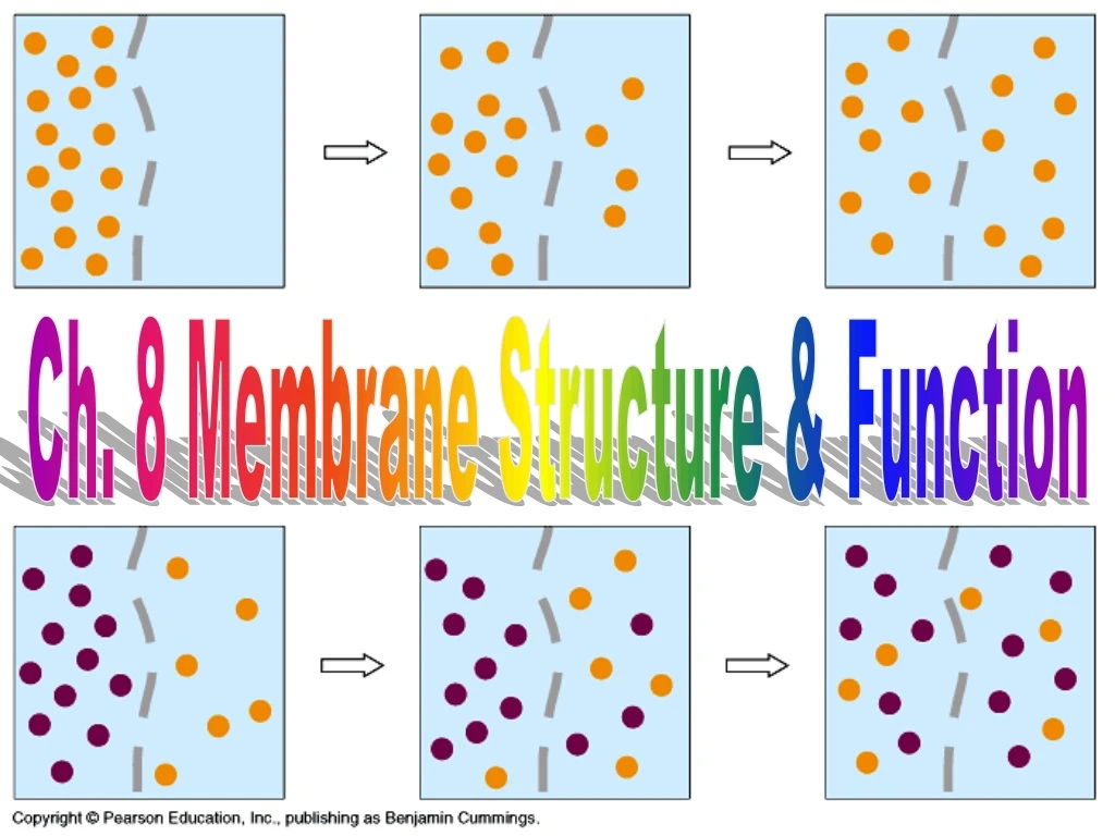
Passive Transport • Passive transport does not require energy from the cell • Diffusion – the tendency for molecules of any substance to spread out into the available space • Substances always diffuse down its concentration gradient – from an area of greater concentration to an area of lesser concentration • Diffusion is spontaneous because it decreases free energy
Osmosis • Osmosis is the passive transport (diffusion) of water across a selectively permeable membrane • Hypertonic– the solution with a higher concentration of solutes • Hypotonic – the solution with a lower concentration of solutes • Isotonic – solutions of equal solute concentration
Water Balance Within Cells • Osmoregulation is the control of water balance within living cells • Animal cells do not have cell walls • If in a hypotonic solution, the cell takes on water quickly and can burst (lyse) • If in a hypertonic solution, the cell loses water and shrivels usually leading to death • Cells usually live in an isotonic environment
Water Balance Within Cells • Paramecium have a contractile vacuole to get rid of excess water
Figure 8.13 The contractile vacuole of Paramecium: an evolutionary adaptation for osmoregulation
Water Balance Within Cells • Plant, prokaryote, fungi, and some protist cells dohave cell walls • If in a hypotonicsolution, the cell takes on water, but the cell wall prevents bursting • Turgor pressure keeps plants upright • If in a hypertonic solution, the cell loses water and pulls away from the cell wall • Plasmolysis usually kills the cell • In an isotonic environment, water does not enter the cell • Causes wilting (cells become flaccid)
Facilitated Diffusion • Use of transport proteins to move substances across the plasma membrane This transport protein alternates between two shapes to move substances through A channel protein allows substance to pass through
Some Functions of Membrane Proteins Video Transport Proteins
Active Transport • The cell must expend energy (ATP) to move substance against the concentration gradient (from low concentration to high concentration)
Figure 8.15 The sodium-potassium pump: a specific case of active transport
Exocytosis • Secretion of macromolecules by the fusion of vesicles with the plasma membrane • A transport vesicle that has budded from the Golgi apparatus moves to the plasma membrane • When it reaches the membrane, the vesicle fuses with it and releases its contents to the outside of the cell
Endocytosis • The cell takes in macromolecules and particulate matter by forming new vesicles from the plasma membrane • Three types • Phagocytosis – the cell engulfs large particles by forming a vacuole from the cell membrane; the vacuole fuses with a lysosome to be digested • Pinocytosis – droplets of fluid are taken into the cell
Receptor-Mediated Endocytosis • The movement of specific molecules into a cell by the inward budding of vesicles containing proteins with receptor sites specific to the molecules being taken in • This enables a cell to acquire bulk quantities of specific substances.