Download
1 / 63
630 likes | 745 Vues
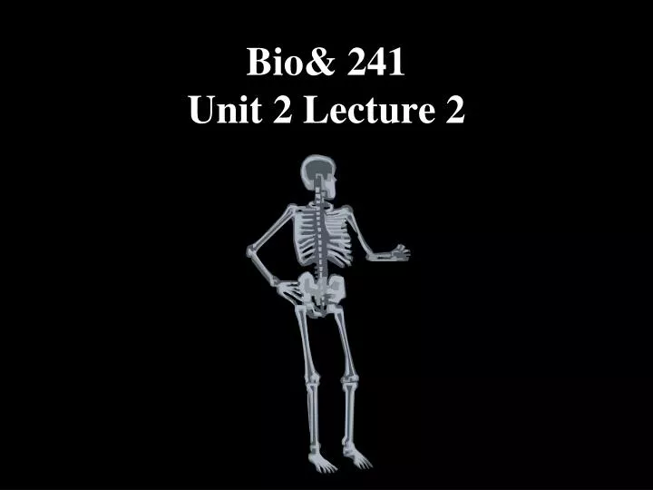
Bio& 241 Unit 2 Lecture 2. CONSISTS OF FOUR TYPES OF CONNECTIVE TISSUE (CT): Cartilage 2. Bone 3. Bone Marrow 4. Periosteum [ Osseous (bone) tissue makes up MOST of the skeleton .]. Bone Tissue: Supportive Connective Tissue . Remember “CT” is composed of: Cells

E N D
CONSISTS OF FOUR TYPES OF CONNECTIVE TISSUE (CT): • Cartilage 2. Bone 3. Bone Marrow 4. Periosteum [Osseous (bone) tissue makes up MOST of the skeleton.] Bone Tissue: Supportive Connective Tissue Remember “CT” is composed of: Cells Extracellular Matrix
Bone Tissue: Supportive Connective Tissue Extracellular Matrix 25% Water 25% Protein or organic matrix 95% Collagen Fibers 5% Chondroitin Sulfate 50% Crystalized Mineral Salts Hydroxyapatite (Calcium Phosphate crystals) Other substances: Lead, Gold, Strontium, Plutonium, can be incorporated in etc. RATIO OF ORGANIC TO NON ORGANIC MATRIX: Youth = 1:1, 50%:50% Adult = 1:2, 33%:66% Elderly = 1:3, 25%:75% (bones become more brittle as we age).
Two Kinds of Bone Compact Bone: 1. Consists of osteons with very little space between them. 2. Composes bone tissue of the diaphysis. FX = Protect and support Spongy Bone: Does NOT contain osteons. Consist of trabeculae Found in short, flat and irregular bones and in the epiphyses of long bones. FX = store RED marrow.
Compact Bone • Compact bone is arranged in units called osteons or Haversian systems. • Osteons (Haversian canal) contain blood vessels, lymphatic vessels, nerves • Surrounding this canal are concentric rings of osteocytes along with the calcified matrix. • Osteons are aligned in the same direction along lines of stress. These lines can slowly change as the stresses on the bone changes.
Histology of Compact Bone • Osteon is concentric rings (lamellae) of calcified matrix surrounding a vertically oriented blood vessel • Osteocytes are found in spaces called lacunae • Osteocytes communicate through canaliculi filled with extracellular fluid that connect one cell to the next cell • Interstitial lamellae represent older osteons that have been partially removed during tissue remodeling
The Trabeculae of Spongy Bone • Latticework of thin plates of bone called trabeculae oriented along lines of stress • Spaces in between these struts are filled with red marrow where blood cells develop • Found in ends of long bones and inside flat bones such as the hipbones, sternum, sides of skull, and ribs. No true Osteons.
BONE FORMATION • All embryonic connective tissue begins as mesenchyme. • Bone formation is termed osteogenesis or ossification and begins when mesenchymal cells provide the template for subsequent ossification. • Two types of ossification occur. • Intramembranous ossification is the formation of bone directly from or within fibrous connective tissue membranes. • Endochondrial ossification is the formation of bone from hyaline cartilage models.
Two Kinds of Ossification 1. Intramembranous Ossification 2. Endochondral Ossification
Intramembranous Ossification Also called dermal ossification because it normally occurs in the deeper layers of connective tissue of the dermis of the skin. All roofing bones of the Skull Frontal bone Parietal bones Occipital bone Temporal bones Mandible Clavicle
Centers of Ossification Centers of Ossification
Endochondral Ossification • All roofing bones of the Skull • Mandible • Clavicle Developing bones are deposited as a hyaline cartilage model and then this cartilage is replaced by bone tissue. All bones of the body except:
Growth at epiphyseal plates Zones of epiphyseal plates Zone of Resting Cartilage Zone of Proliferating Cartilage Zone of Hypertrophic Cartilage Zone of Calcified Cartilage
Zones of Growth in Epiphyseal Plate • Zone of resting cartilage • anchors growth plate to bone • Zone of proliferating cartilage • rapid cell division (stacked coins) • Zone of hypertrophic cartilage • cells enlarged & remain in columns • Zone of calcified cartilage • thin zone, cells mostly dead since matrix calcified • osteoclasts removing matrix • osteoblasts & capillaries move in to create bone over calcified cartilage
Growth at epiphyseal plates Zones of epiphyseal plates Zone of Resting Cartilage Zone of Proliferating Cartilage Zone of Hypertrophic Cartilage Zone of Calcified Cartilage
Growth at epiphyseal plates Zones of epiphyseal plates Zone of Resting Cartilage Zone of Proliferating Cartilage Zone of Hypertrophic Cartilage Zone of Calcified Cartilage
Growth at epiphyseal plates Zones of epiphyseal plates Zone of Resting Cartilage Zone of Proliferating Cartilage Zone of Hypertrophic Cartilage Zone of Calcified Cartilage
Growth at epiphyseal plates Zones of epiphyseal plates Zone of Resting Cartilage Zone of Proliferating Cartilage Zone of Hypertrophic Cartilage Zone of Calcified Cartilage
Growth in Thickness • Bone can grow in thickness or diameter only by appositional growth. • The steps in these process are: • Periosteal cells differentiate into osteoblasts which secrete collagen fibers and organic molecules to form the matrix. • Ridges fuse and the periosteum becomes the endosteum. • New concentric lamellae are formed. • Osetoblasts under the peritsteum form new circumferential lamellae.
Bone Growth in Width • Only by appositional growth at the bone’s surface • Periosteal cells differentiate into osteoblasts and form bony ridges and then a tunnel around periosteal blood vessel. • Concentric lamellae fill in the tunnel to form an osteon.
Factors That Affect Bone Growth Minerals Vitamins Hormones Exercise
Factors That Affect Bone Growth Minerals Calcium Makes bone matrix hard Hypocalcemia: low blood calcium levels. Hypercalcemia: high blood calcium levels.
Factors That Affect Bone Growth Vitamins Vitamin A Controls activity, distribution, and coordination of osteoblasts/osteoclasts Vitamin B12 May inhibit osteoblast activity Vitamin C Helps maintain bone matrix, deficiency leads to decreased collagen production which inhibits bone growth and repair (scury) disorder due to a lack of Vitamin C Vitamin D (Calcitriol) Helps build bone by increasing calcium absorption. Deficiencies result in “Rickets” in children
Factors That Affect Bone Growth Hormones Human Growth Hormone Promotes general growth of all body tissue and normal growth in children Insulin-like Growth Factor Stimulates uptake of amino acids and protein synthesis Insulin Promotes normal bone growth and maturity Thyroid Hormones Promotes normal bone growth and maturity Estrogen and Increases osteogenesis at puberty Testosterone and is responsible for gender differences of skeletons
Bone Fractures Closed fracture:one that does not produce an open wound in the skin Open fracture: one in which a wound through the adjacent or overlying soft tissues communicates with the site of the break. Compound fracture: A fracture in which the bone is sticking through the skin. Also has been called an open fracture. Simple fracture : an uncomplicated fracture in which the broken bones to not pierce the skin. Also has been called a closed fracture. Comminuted fracture: The bone is splintered or crushed, Can be viewed as a “closed compound fracture”
Bone Fractures Colles' fracture: fracture of the lower end of the radius, the lower fragment being displaced backward Greenstick fracture: one side of a bone is broken, the other being bent. Most common in children. Impacted fracture one bone fragment is firmly driven into the other. Common with vertebra. Pathologic fracture: due to weakening of the bone structure by pathologic processes, such as neoplasia, osteomalacia, or osteomyelitis Pott's fracture: fracture of the lower part of the fibula, with serious injury of the lower tibial articulation, usually a chipping off of a portion of the medial malleolus, or rupture of the medial ligament
Bone Fractures Terms: Partial/Complete Displaced/Non-displaced Other Fractures: Spiral Transverse Longitudinal Pathologic
Dislocations Subluxation : an incomplete or partial dislocation of a joint or organ. Luxation: a complete dislocation of A joint or organ.
Steps in Fracture Repair 1. Formation of a fracture hematoma Immediately after the fracture, there is a sharp fracture line with associated soft tissue swelling. At the fracture Site, there is abundant hematoma with beginning fibroblastic penetration.
Steps in Fracture Repair • Fibrocartilaginous Callus • Formation At 2 weeks there is much visible callus. There is also bone resorption and osteoporosis, both difficult to see in this case because of the overlying callus. There has been migration of chondroblasts into the area and cartilage is beginning to cover the ends of the fracture. New osteous tissue is produced enchondrally.
Steps in Fracture Repair 3. Bony Callus Formation At 2 months, bony callus with sharp margins bridges the fracture and the fracture line itself begins to disappear.
Steps in Fracture Repair 4. Bone Remodeling At 5-6 months, the marrow cavity is continuous and the compact bone of the diaphysis has been reformed.
Congenital Skeletal Birth Defects Congenital TalipesEquinovarus (CTEV),[ occurs in about one in every 1,000 live births. Approximately 50% of cases of clubfoot are bilateral Occurs in males more often than in females by a ratio of 2:1.