Download
1 / 37
400 likes | 664 Vues
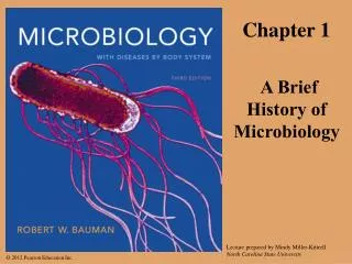
chapter one: the history of microbiology. microbes. microscopic (small) organisms, viruses, prions. scientific names. Staphylococcus aureus and Escherichia coli can be part of the normal flora of the human body; S. aureus is found on the skin and E. coli in the large intestine.

E N D
microbes microscopic (small) organisms, viruses, prions
scientific names Staphylococcus aureus and Escherichia coli can be part of the normal flora of the human body; S. aureus is found on the skin and E. coli in the large intestine.
microbes and human welfare • microbial ecology • biogeochemical & nutrient cycling • bioremediation • degrade organic matter & pollutants • biological insecticides • microbes selectively pathogenic to insects • biotechnology • microbes producing food &chemicals • recombinant DNA technology • production of vaccines, proteins, et al. • gene therapy • genetically modified bacteria protectcrops from insects & freezing
human bacteria • normal microbiota • those present in & on the human body • prevent growth of pathogens • produce growth factors (folic acid & vitamin K) • produce antimicrobial compounds • pathogenic organisms
chapter three, part I: microscopyµscopy lab information
resolution • resolution: the ability to distinguish two points • shorter = greater resolution (electron > light) unaided eye light microscope electron microscope
light microscopy • simple vs compound- # of lenses • compound microscope- imagemagnified by objective& ocular lenses • total magnification =objective lens ocular lens
microscopy INCREASING RESOLUTION
biogenesis or spontaneous generation 1768 Spallanzani biogenesis 1745: John Needham spontaneous generation
Chapter One Learning Objectives • What types of “organisms” are studied in microbiology? Which of these are considered non-living? • How are scientific names correctly written? • Six specific microbiological breakthroughs from the “Golden Age of Microbiology” were discussed in class. For each, you should commit to memory the date, researcher and contribution to the field of microbiology. • Discuss the four major experiments explained in class that led to the refutation of Spontaneous Generation and the scientific acceptance of the theory of Biogenesis. For each leading up to the work of Louis Pasteur, why did the scientific community as a whole remain unconvinced of the novel theory of Biogenesis. • Understand how microorganisms can play both a beneficial and a detrimental role in their interactions with people. Include normal flora in your discussion.
the prokaryotic cell: external structures • glycocalyx: capsule or slime layer • attachment • prevent phagocytosis • flagella • motility • fimbriae • attachment • pili • DNA transfer
axial filaments/endoflagella: spirochetes anchored at one end; rotate to cause movement
laboratory culture & aseptic technique • liquid & solid media are either complex or chemically defined • solid media have agar
(enteric peritrichous) taxis • small size means they sense gradients temporally, not spatially • attractants = tumble • repellants = tumble
the cell wall • prevents osmotic lysis; usually peptidoglycan • Mycoplasmas: lack cell walls, sterols in plasma membrane • Archaea: wall-less/pseudomurein (no NAM & D-amino acids)
acid-fast (AFB) cell walls waxy lipid (mycolic acid) bound to peptidoglycan Mycobacterium (Nocardia)
ester vs. ether linkages:the archaean cell membrane ester linkage ether linkage
inclusions • metachromatic granules • phosphate reserves • energy reserves • polysaccharide granules • lipid inclusions • sulfur granules • carboxysomes • CO2 fixation • gas vacuoles • buoyancy • magnetosomes • iron oxide (destroys H2O2)
sporulation 8–10 hours, 113 genes • chromosome condensation • septumformation • foresporeengulfed • peptidoglycancortex formed • spore coatformed • resistance mechanisms develop, endospore released
Chapter Four Learning Objectives • Identify and correctly name the five morphological types as well as the various arrangements of bacterial cells discussed in lecture. • Understand the location and function of each of the following prokaryotic structures: glycocalyx, capsule, slime layer, flagella, fimbriae, pili, cell wall, cell membrane, nucleoid region and inclusions. • Identify and correctly name the four flagellar arrangements. • Discuss the basic mechanism for bacterial motility. Include in your discussion how bacterial flagella differ from eukaryotic flagella in structure and motility and how different bacterial arrangements will affect how bacterial cells move. Include endoflagella/axial filaments in your discussion. • Discuss positive and negative chemotaxis and phototaxis. How do bacterial cells respond to attractants and repellants? • Why is the cell wall a necessary component of most bacterial cells? How is the cell wall of a bacterium different from plants, fungi, Archaea and Mycoplasmas? • Understand the chemical make-up of Gram positive, Gram negative and Acid Fast cell walls. • What are the major structural differences between bacterial and archaeal cell membranes? • Provide the name and function of each of the bacterial inclusions discussed in lecture. • How does a nucleoid region differ from a nucleus? How is it similar? • Discuss the process of sporulation and germination. For what is a spore used? Can all bacteria produce spores?
staining increases resolution • stains increase visibility & emphasizes structures • positive (basic) stains • absorbed by cell • chromophore is cation • negative (acidic) stains • repelled by cell • chromophore is anion • heat-fixed bacterial smears • attach to slide & kill • simple stain • differential stains: distinguish Bacteria • stain & counterstain
differential & special staining • Differential Staining • Gram stain • Abx treatment, diagnosis,characterization • Acid-Fast stain • diagnosis: TB • Spore stain • food preservation& safety • Special staining • Capsule • pathogenesis
Chapter Three Learning Objectives • Define resolution. How does wavelength relate to resolving power? • Understand the mechanism by which light microscopes, scanning electron microscopes and transmission electron microscope allow you to visualize an image. Commit to memory the resolving power of each. • Explain the use of positive/basic stains and negative/acidic stains. How does each work and what part of the cell does each stain? • Define differential staining. For each technique discussed in class, understand the mechanism of action and what “positive” and “negative” look like.