Download
1 / 75
770 likes | 1.09k Vues
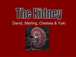
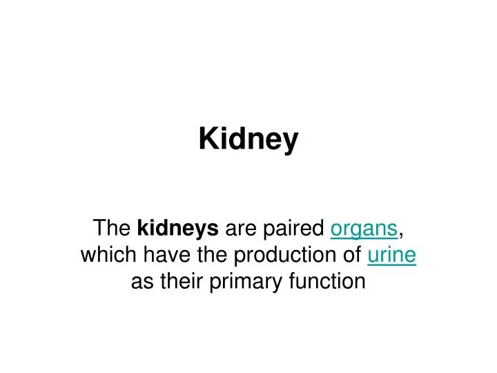
Kidney. The kidneys are paired organs , which have the production of urine as their primary function. The two kidneys are bean-shaped organs located near the middle of the back, just below the rib cage to the left and right of the spine.

E N D
Kidney The kidneys are paired organs, which have the production of urine as their primary function
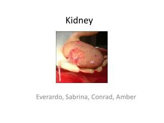
The two kidneys are bean-shaped organs located near the middle of the back, just below the rib cage to the left and right of the spine. • Each about the size of a fist, these organs act as sophisticated filters for the body. • They process about 200 quarts of blood a day to sift out about 2 quarts of waste products and extra water that eventually leave the body as urine.
Blood enters the kidneys through arteries that branch inside the kidneys into tiny clusters of looping blood vessels. • Each cluster is called a glomerulus, which comes from the Greek word meaning filter. • The plural form of the word is glomeruli. • There are approximately 1 million glomeruli, or filters, in each kidney. • The glomerulus is attached to the opening of a small fluid-collecting tube called a tubule. • Blood is filtered in the glomerulus, and extra water and wastes pass into the tubule and become urine. • Eventually, the urine drains from the kidneys into the bladder through larger tubes called ureters.
secondary functions concerned with homeostatic functions. • These include the regulation of electrolytes, acid-base balance, and blood pressure. • In producing urine, the kidneys excrete wastes such as urea and ammonium; • The kidneys also are responsible for the reabsorption of glucose and amino acids. • Finally, the kidneys are important in the production of hormones including vitamin D, renin and erythropoietin.
Located behind the abdominal cavity in the retroperitoneum, the kidneys receive blood from the paired renal arteries, and drain into the paired renal veins. • Each kidney excretes urine into a ureter, itself a paired structure that empties into the urinary bladder.
The substance, or parenchyma, of the kidney is divided into two major structures: superficial is the renal cortex and deep is the renal medulla. • Grossly, these structures take the shape of 8 to 18 cone-shaped renal lobes, each containing renal cortex surrounding a portion of medulla called a renal pyramid (of Malphigi). • Between the renal pyramids are projections of cortex called renal column
Renal physiology is the study of kidney function, while nephrology is the medical specialty concerned with diseases of the nephron, which is the functional unit of the kidney.
Glomerular Diseases • Many diseases affect kidney function by attacking the glomeruli, the tiny units within the kidney where blood is cleaned. • Glomerular diseases include many conditions with a variety of genetic and environmental causes, but they fall into two major categories: • Glomerulonephritis (gloh-MEHR-yoo-loh-nef-RY-tis) • Describes the inflammation of the membrane tissue in the kidney that serves as a filter, separating wastes and extra fluid from the blood. • Glomerulosclerosis (gloh-MEHR-yoo-loh-skleh-ROH-sis) • Describes the scarring or hardening of the tiny blood vessels within the kidney. • Although glomerulonephritis and glomerulosclerosis have different causes, they can both lead to kidney failure.
Each glomerulus-and-tubule unit is called a nephron. • Each kidney is composed of about 1 million nephrons. • In healthy nephrons, the glomerular membrane that separates the blood vessel from the tubule allows waste products and extra water to pass into the tubule while keeping blood cells and protein in the bloodstream.
Glomerular diseases damage the glomeruli, letting protein and sometimes red blood cells leak into the urine. • Sometimes a glomerular disease also interferes with the clearance of waste products by the kidney, so they begin to build up in the blood.
Furthermore, loss of blood proteins like albumin in the urine can result in a fall in their level in the bloodstream. • In normal blood, albumin acts like a sponge, drawing extra fluid from the body into the bloodstream, where it remains until the kidneys remove it. • But when albumin leaks into the urine, the blood loses its capacity to absorb extra fluid from the body. • Fluid can accumulate outside the circulatory system in the face, hands, feet, or ankles and cause swelling. • [Top]
Symptoms of glomerular disease • proteinuria: large amounts of protein in the urine • hematuria: blood in the urine • reduced glomerular filtration rate: inefficient filtering of wastes from the blood • hypoproteinemia: low blood protein • edema: swelling in parts of the body
some of these symptoms have signs, or visible manifestations: • Proteinuria may cause foamy urine. • Blood may cause the urine to be pink or cola-colored. • Edema may be obvious in hands and ankles, especially at the end of the day, or around the eyes when awakening in the morning, for example.
DIAGNOSIS • URINE ANALYSIS- • protein in the urine- • which may be referred to as “nephrotic range” if levels are very high. • Red blood cells in the urine • are a frequent finding as well, particularly in some forms of glomerular disease . • Urinalysis provides information about kidney damage by indicating levels of protein and red blood cells in the urine.
Blood tests-measure the levels of waste products such as • creatinine • urea nitrogen • To determine whether the filtering capacity of the kidneys is impaired. • If these lab tests indicate kidney damage, • ultrasound or an x ray to see whether the shape or size of the kidneys is abnormal. • These tests are called renal imaging.
But since glomerular disease causes problems at the cellular level. • kidney biopsy—a procedure in which a needle is used to extract small pieces of tissue for examination with different types of microscopes, each of which shows a different aspect of the tissue. • A biopsy may be helpful in confirming glomerular disease and identifying the cause.
causes glomerular disease- • A number of different diseases can result in glomerular disease. • It may be the direct result of an infection or a drug toxic to the kidneys, • or it may result from a disease that affects the entire body, like diabetes or lupus. • Many different kinds of diseases can cause swelling or scarring of the nephron or glomerulus. • Sometimes glomerular disease is idiopathic, meaning that it occurs without an apparent associated disease.
can overlap: • that is, a disease might belong to two or more of the categories. • For example, diabetic nephropathy is a form of glomerular disease that can be placed in two categories: • systemic diseases, since diabetes itself is a systemic disease, • and sclerotic diseases, because the specific damage done to the kidneys is associated with scarring.
Proximal tubule • The proximal tubule is the portion of the duct system of the nephron leading from Bowman's capsule to the loop of Henle. • The most distinctive characteristic of the proximal tubule is its brush border (or "striated border"). • The luminal surface of the epithelial cells of this segment of the nephron is covered with densely packed microvilli forming a border readily visible under the light microscope. • The microvilli greatly increase the luminal surface area of the cells, presumably facilitating their resorptive function.
The proximal tubule as a part of the nephron can be divided into two sections, • pars convoluta • pars recta. • Differences in cell outlines exist between these segments, and therefore presumably in function too.
Pars convoluta • The "Pars convoluta" is the initial convoluted portion. • In relation to the morphology of the kidney as a whole, the convoluted segments of the proximal tubules are confined entirely to the renal cortex.
Pars recta • The "Pars recta" is the following straight (descending) portion. • Straight segments descend into the outer medulla. • Secretion • Many types of medications are secreted in the proximal tubule
Substance-salt and water -salt and waterapproximately two-thirds. • organic solutes (primarily glucose and amino acids)-100% • Potassium-65% • Urea-50% • Phasphate-80%
Pathophysiology in kidney disease • Proximal tubular epithelial cells (PTECs) have a pivotal role in kidney disease. • Malignant • Most renal cell carcinoma, the most common form of kidney cancer, arises from the convoluted tubules.
Non-malignant • Acute tubular necrosis occurs when PTECs are directly damaged by toxins such as • Antibiotics (e.g. gentamicin), • Pigments (e.g. myoglobin and sepsis (e.g. mediated by lipopolysaccharide from gram negative bacteria). • Renal tubular acidosis (proximal type) (Fanconi syndrome) occurs when the PTECs are unable to properly reabsorb glomerular filtrate so that there is increased loss of bicarbonate, glucose, amino acids and phosphate.
Diseases of the kidney are diverse, but individuals with kidney disease frequently display characteristic clinical features. Common clinical presentations include the nephritic and nephrotic syndromes, • acute kidney failure, • chronic kidney disease, • urinary tract infection, nephrolithiasis, and urinary tract obstruction.
Nephritic syndrome (or acute nephritic syndrome) • Is a collection of signs (known as a syndrome) associated with disorders affecting the kidneys, more specifically glomerular disorders. • It's characterized by having small pores in the podocytes of the glomerulus, large enough to permit proteins (proteinuria) and red blood cells (hematuria) to pass into the urine. • By contrast, nephrotic syndrome is characterized by only proteins (proteinuria) moving into the urine. • Both nephritic syndrome and nephrotic syndrome result in hypoalbuminemia due to protein mainly albumin moving from the blood to the urine.
Hematuria can be caused by bleeding anywhere in the urinary tract, but if the RBCs are trapped in urinary casts, it is more likely that the bleeding originated in the nephron, and Nephritic syndrome becomes more probable .
Signs and symptoms • Nephritic syndrome is characterized by; • hematuria (blood in the urine), with red blood cell (RBC) casts present in the urine • proteinuria (protein in the urine) - small amounts of protein are lost in the urine, but this is usually trivial (<3.5 g/day) • hypertension (high blood pressure)- mild • uremia - due to retention of waste products
and variable renal insufficiency, with; • azotemia (elevated blood nitrogen) • oliguria (low urine output <400 mL/day) • The main features are hypertension and RBC casts. The proteinuria in nephritic syndrome is not usually severe, but may occasionally be heavy enough to be in the range usually found in nephrotic syndrome. • Mnemonic: PHAROH = Proteinuria, Hematuria, Azotemia, RBC casts, Oliguria, Hypertension.
Nephrotic syndrome (NS), also known as nephrosis, is defined by the presence of- • Nephrotic-range proteinuria, • Edema, • Hyperlipidemia. • Hypoalbuminemia. • Nephrotic-range proteinuria in adults is characterized by protein excretion of 3.5 g or more per day.
Acute Renal Failure • Acute renal failure (ARF) is the rapid breakdown of renal (kidney) function that occurs when high levels of uremic toxins (waste products of the body's metabolism) accumulate in the blood. • ARF occurs when the kidneys are unable to excrete (discharge) the daily load of toxins in the urine
Based on the amount of urine that is excreted over a 24-hour period, patients with ARF are separated into two groups: • Oliguric: patients who excrete less than 500 milliliters per day (< 16 oz/day) • Nonoliguric: patients who excrete more than 500 milliliters per day (> 16 oz/day)
In nonoliguric patients, the urine is of poor quality (i.e., contains little waste) because the blood is not well filtered, despite the fact that an adequate volume of urine is excreted. • Both kidneys are failing when ARF occurs. • One normally functioning kidney can maintain adequate blood filtering.
Definition • Chronic kidney failure is a gradual loss of your kidneys' filtering ability, usually due to high blood pressure or diabetes. • When kidney function is seriously impaired, dangerous levels of fluid and waste can quickly accumulate in your body. • In the early stages of chronic kidney failure, you may have few signs or symptoms. • Many people with chronic kidney failure don't realize they have a problem until their kidney function has decreased to less than 25 percent of normal.
The main goal of treatment of chronic kidney failure is to halt or delay progression of the disease, usually by controlling the underlying cause. • Chronic kidney failure can progress to end-stage kidney disease, which is fatal without artificial filtering (dialysis) or a kidney transplant.
Symptoms • Signs and symptoms may include some or many of the following: • High blood pressure • Decreased urine output or no urine output • Darkly colored urine • Anemia • Nausea or vomiting
Loss of appetite • Sudden weight change • A general sense of discomfort and unease (malaise) • Fatigue and weakness • Headaches that seem unrelated to any other cause • Sleep problems • Decreased mental sharpness
Definition of Pyelonephritis • Pyelonephritis is a serious bacterial infection of the kidney that can be acute or chronic. • It presents with dysuria (painful voiding of urine), abdominal pain (radiating to the back on the affected side) • and tenderness of the bladder area and the side of the involved kidney (costovertebral angle tenderness) which may be elicited by performing the kidney punch
In many cases there are systemic symptoms in the form of fever, rigors (violent shivering while the temperature rises), headache, and vomiting. • In severe cases, delirium may be present. • Severe cases of pyelonephritis lead to sepsis, a systemic response to infection characterized by fever, a raised heart rate, rapid breathing and decreased blood pressure (occasionally leading to septic shock).
Causes • Most cases of "community-acquired" pyelonephritis are due to bowel organisms that enter the urinary tract. • Common organisms are E. coli (70-80%) and Enterococcus faecalis. • Hospital-acquired infections may be due to coliforms and enterococci, as well as other organisms uncommon in the community (e.g. Klebsiella spp., Pseudomonas aeruginosa). • Most cases of pyelonephritis start off as lower urinary tract infections, mainly cystitis and prostatitis.
Pathology • Acute pyelonephritis is an exudative purulent localized inflammation of the renal pelvis (collecting system) and kidney. • The renal parenchyma presents in the interstitium abscesses (suppurative necrosis), consisting in purulent exudate (pus): neutrophils, fibrin, cell debris and central germ colonies (hematoxylinophils). • Tubules are damaged by exudate and may contain neutrophil casts. In the early stages, glomeruli and vessels are normal.