Download
1 / 16
190 likes | 475 Vues
Kidney. Chapter 11.3. 11.3.1 Define excretion. Excretion : Method of removing waste products (such as urea). Outline the need for excretion in all living organisms. Keeps the by-products of metabolism from building up and poisoning the body fluids

E N D
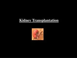
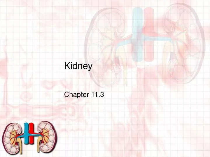
Kidney Chapter 11.3
11.3.1 Define excretion. • Excretion: Method of removing waste products (such as urea)
Outline the need for excretion in all living organisms. • Keeps the by-products of metabolism from building up and poisoning the body fluids • Disposing of metabolic wastes by moving solutes (and therefore, water) between an animal’s internal fluids and the external environment
Describe and show the relationship among the processes of filtration, secretion, and reabsorption. • Filtration: Blood is filtered • Proteins and other large molecules stay in blood. • Water and small solutes are sent to the excretory system. This creates filtrate • Reabsorption: Filtrate has water and other ions (glucose, salts, amino acids) that the body still needs. As a result, these substances are taken back through active or passive means. The water and ions move into the tissue fluid or into the neighboring capillaries/arterioles • Secretion: Opposite of reabsorption. • Molecules are moved back into the filtrate (hormones, hydrogen ions)
Using a diagram, identify and give the function of each structure in the mammalian excretory system. • Blood Vessels: Bring/return blood • Bladder: Store urine • Kidney: Produces urine • Ureter: Carries urine to the bladder • Urethra: Conducts urine from bladder to outside • Diagram to be done in class.
11.3.2 Draw and label the structure of the kidney. • Cortex: Site of Bowman’s capsule. Solute secretion and reabsorption • Medulla: Loop of Henle. Water and salt balance. • Ureter: Tube for transport of urine to the urinary bladder • Renal Blood Vessels: Renal artery (into kidney), renal vein (away from kidney) • Diagram to be done in class The kidney
11.3.3 Annotate a diagram of a glomerulus and associated nephron to show the function of each part. • Nephron is the functional unit of the kidney which consists of a single long tube and ball of capillaries called the glomerulus • The tube dead ends as Bowman’s capsule. Bowman’s capsule surrounds the glomerulus. • Diagram to be done in class. Glomerulus and Nephron
Outline the role of the kidney in excretion and the maintenance of water balance. • Nephrons do the work in the kidney by regulating blood composition and excreting wastes. • The blood is first filtered, water and solutes are forced into the nephron creating filtrate. • The filtrate is modified by reabsorbing water and solutes, or by secreting them as urine is produced. • Depending on the body’s salt and water balance, the modification can be controlled to retain or release more or less water/solutes. • Antidiuretic hormone is the primary means of control. (More ADH=more water retained)
11.3.4 Explain the process of ultrafiltration including blood pressure, fenestrated blood capillaries, and basement membrane. • Ultrafiltration: Production of filtrate from the blood. Blood enters the capillaries of the glomerulus, and water/solutes are forced into Bowman’s capsule to create the filtrate. • Blood pressure: HIGH! The exiting vessel is smaller than the entering vessel. This forces the molecules through
11.3.4 Explain the process of ultrafiltration including blood pressure, fenestrated blood capillaries, and basement membrane. • Fenestrated capillaries: Means it has pores/slits. • Basement Membrane: Gel of glycoproteins. Prevents large proteins through. • Diagram to be done in class. Filtration
11.3.5 Define osmoregulation. • Osmoregulation: how animals regulate solute balance and the gain and loss of water • Osmoregulators: animals that regulate the internal osmolarity controlling water intake/release • Marine animals, freshwater animals, terrestrial animals and humans • Osmoconformers: animals that do not actively adjust their internal osmolarity. Allow their solute concentration to change with the environment • Most marine invertebrates
11.3.6 Explain the reabsorption of glucose, water, and salts in the proximal convoluted tubule including the roles of microvilli, osmosis and active transport. • Selective reabsorption allows the body to take back 90% of the filtrate (such as glucose, Na+, K+, Mg2+, Ca2+, Cl-). • Microvilli line the inside of the tubule, increasing the surface area for absorption. • Active transport pumps most of the ions (except Cl-) across the membrane, increasing solute concentration of the tubule cells. • Osmosis occurs when water follows the solutes. 80% of the water in the filtrate is re-absorbed.
11.3.7 Explain the roles of the loop of Henle, medulla, collecting duct, and ADH in maintaining the water balance of the blood. • Remember that the nephron has three turns. The first turn (Loop of Henle) moves deep into the kidney’s medulla. • Descending limb-moving towards medulla • water permeable • Ascending limb-moving out from medulla • non-water permeable • pump out sodium, making medulla salty • As a result, the ‘salty’ medulla will draw out water from the descending limb.
11.3.7 Explain the roles of the loop of Henle, medulla, collecting duct, and ADH in maintaining the water balance of the blood. • Overall, the filtrate ends up less concentrated after leaving the ascending limb • Collecting duct: where the urine collects at the end of the nephron • If water levels are too low, ADH is released, making the collecting ducts permeable to water, concentrating the urine. • If water levels are too high, no ADH is released, water stays in the urine and it is dilute.
11.3.8 Explain the differences in the concentration of proteins, glucose, and urea between blood plasma, glomerular filtrate, and urine. • Proteins are too large to fit through basement membrane so don’t become part of filtrate or urine. • Glucose becomes part of filtrate but active transport takes 100% of glucose back into capillary bed. • Urea is not toxic in low levels in blood plasma. High concentration of urea in urine is due to reabsorption of water.
11.3.9 Explain the presence of glucose in the urine of untreated diabetic patients. • Untreated Diabetes = high blood glucose • Recall: Glucose becomes part of filtrate but active transport takes 100% of glucose back into capillary bed • If plasma glucose levels are too high, active transport can’t keep up and can’t move all glucose back into bloodstream SO, some glucose remains in the urine