Download
1 / 43
490 likes | 1.22k Vues
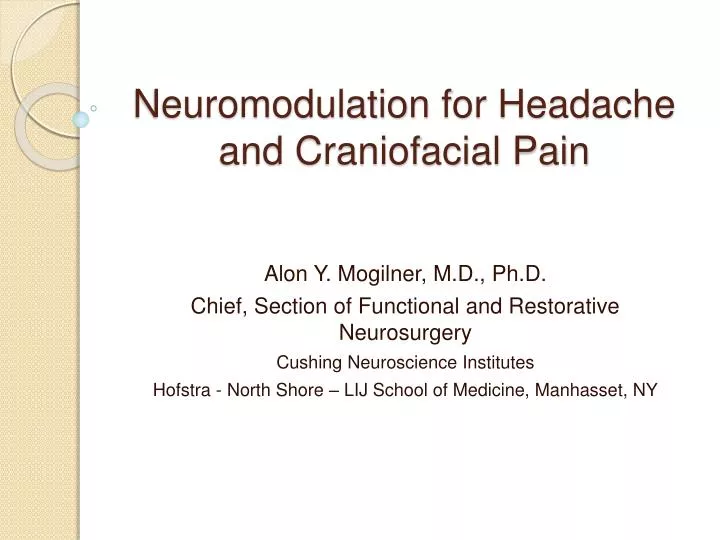
Neuromodulation for Headache and Craniofacial Pain. Alon Y. Mogilner, M.D., Ph.D. Chief, Section of Functional and Restorative Neurosurgery Cushing Neuroscience Institutes Hofstra - North Shore – LIJ School of Medicine, Manhasset, NY. Disclosures.

E N D
Neuromodulation for Headache and Craniofacial Pain Alon Y. Mogilner, M.D., Ph.D. Chief, Section of Functional and Restorative Neurosurgery Cushing Neuroscience Institutes Hofstra - North Shore – LIJ School of Medicine, Manhasset, NY
Disclosures • Alon Y. Mogilner, M.D., Ph.D. receives honoraria and grant support from Medtronic, and grant support from St. Jude Medical • Peripheral Neuromodulation for Headache and Craniofacial Pain is not FDA approved, and thus represents an off-label indication.
Introduction • Neurosurgical management of chronic headache and craniofacial pain syndromes has been an evolving concept over the past 50 years • Early interest in neurostimulation, for these conditions, focused on deep brain targets • Hosobuchi, Adams, and Rutkin, thalamic stimulation to treat facial anesthesia dolorosa, 19731 • Mazars and Pull, intermittent stimulation of nVPL to treat intractable headache, 19762 • Hosobuchi Y, Adams JE, Rutkin B. Chronic thalamic stimulation for the control of facial anesthesia dolorosa. Arch Neurol 1973;29(3):158-61. • Mazars G, Pull H. Neurosurgical treatment of headaches. Minerva Med 1976; 67(31):2020-2.
Occipital Nerve Stimulation • Goadsby (1997): Stimulation of greater occipital nerve (GON) in cats resulted in increased metabolic activity of the trigeminal nucleus caudalis and cervical dorsal horn3 • Weiner (1999): Peripheral neurostimulation for occipital neuralgia4 • Goadsby (2004): ONS for migraine headache5 3. Goadsby PJ, Knight YE, Hoskin KL. Stimulation of the greater occipital nerve increases metabolic activity in the trigeminal nucleus caudalis and cervical dorsal horn of the cat. Pain 1997; 73(1):23-8. 4. Weiner RL, Reed KL. Peripheral neurostimulation for control of intractable occipital neuralgia. Neuromodulation 1999; 2(3):217-21. 5. Matharu MS, Bartsch T, Ward N, Frackowiak RSJ, Weiner RL, Goadsby PJ. Central neuromodulation in chronic migraine patients with suboccipital stimulators: A PET study. Brain 2004;127:220-30.
Peripheral Stimulation: headache and facial pain • Most Common technique: • Occipital Nerve Stimulation • Occipital stimulation • “BOTH” stimulation • Other techniques: • Trigeminal branch stimulation • Supraorbital • Supratrochlear • Auriculotemporal
Anatomy • The occipital nerves are derived from C2 • The C2 ventral ramus merges with the cervical plexus and contributes to the lesser occipital nerve • The sensory medial branch of the C2 dorsal ramus contributes to the greater occipital nerve
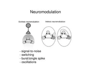
Mechanisms • The mechanism of action of ONS has not been fully elucidated • Afferents from pain-producing cranial structures (dura, blood vessels) likely source of primary headache syndromes • Pain oftentimes is not constrained within trigeminal innervation territories • There is frequently involvement of GON innervation territories
Mechanisms • Role of Trigemino-Cervical Complex? • Afferents from meninges terminate in caudal trigeminal nucleus, in medullary dorsal horn • This nucleus extends down to C2 • Afferents from back of head travel along GON to C2 • Convergent Neurons • Plasticity • This model may not apply though • Mechanism may actually be more similar to peripheral field stimulation (Pain Gate)
Epidermis Dermis Subcutaneous Tissue Electrode Subcutaneous nerves
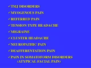
Headache/Facial Pain • Headache • Tension headache • Migraine and equivalents • Non-neurogenic pain • Sinusitis • TMJ pain • Cervicogenic headaches • Occipital neuralgia • Cluster headache • Trigeminal neuralgia • Trigeminal Neuropathic pain • Postsurgical pain • Postherpetic neuralgia • Anesthesia dolorosa • Central Post-Stroke pain • Other cranial neuralgias
Headache/Facial Pain • Headache • Tension headache • Migraine and equivalents • Non-neurogenic pain • Sinusitis • TMJ pain • Cervicogenic headaches • Occipital neuralgia • Cluster headache • Trigeminal neuralgia • Neuropathic pain • Postsurgical pain • Postherpetic neuralgia • Anesthesia dolorosa • Central post-stroke pain • Atypical facial pain
Burchiel Classification of Facial Pain • Trigeminal neuralgia, type 1, (TN1): facial pain of spontaneous onset with greater than 50% limited to the duration of an episode of pain (temporary pain). • Trigeminal neuralgia, type 2, (TN2): facial pain of spontaneous onset with greater than 50% as a constant pain. • Trigeminal neuropathic pain, (TNP): facial pain resulting from unintentional injury to the trigeminal system from facial trauma, oral surgery, ear, nose and throat (ENT) surgery, root injury from posterior fossa or skull base surgery, stroke, etc. • Trigeminal deafferentation pain, (TDP): facial pain in a region of trigeminal numbness resulting from intentional injury to the trigeminal system from neurectomy, gangliolysis, rhizotomy, nucleotomy, tractotomy, or other denervating procedures. • Symptomatic trigeminal neuralgia, (STN): pain resulting from multiple sclerosis. • Postherpetic neuralgia, (PHN):pain resulting from trigeminal Herpes zoster outbreak. (SHINGLES). • Atypical facial pain, (AFP): is facial pain of unknown origin.
Peripheral Stimulation: Procedure • Percutaneous trial performed under local anesthesia • ONS: Supine with head turned lateral position preferred • prone position used in patients who could not be positioned supine/lateral • Lateral or prone position • Trial performed under local anesthesia with sedation (propofol, dexmetetomidine, versed) • Intraoperative testing not always performed • Leads placed horizontal approximately 1 cm above level of C1 arch • May depend on previously placed hardware • OC fusion hardware, • VP shunt hardware
ONS: technique • Cranial and cervical hardware may alter patient positioning, electrode placement
Procedure: Percutaneous Trial • Outpatient trial analogous to SCS trial (4-7 days) • Special considerations: • Chronic migraine • H/A wax and wane with menstrual cycle • Cluster headache • Delay in onset of efficacy • Headache location: • Frontal H/A with no radiation from occipital region • Consider trigeminal branch leads
Procedure: Permanent Implant • Patients usually return with trial leads in place • Leads are removed after induction of general anesthesia • New leads placed and anchored to retromastoid fascia • Extensions rarely used • Generator location: • infraclavicular (most common) • Flank/buttock • abdomen
Complications • Lead migration: most common ONS complication • Surgical technique, anchoring technique not standardized
Complications: Wound Erosion • Be proactive regarding potential wound erosions • Hardware not optimized for peripheral applications
Results: Literature Survey NANS 2009
Neurostimulation for Migraine: Clinical Trials • 3 to date, Boston Scientific, Medtronic, St. Jude • Most recent: St. Jude study • Significant group differences for reduction in number of headache days, MIDAS, Zung PAD, VAS, quality of life and satisfaction at 12 weeks were observed (p< 0.05). • In the Active and Control groups respectively, number of headache days (defined as a > 4 hours duration at moderate intensity) decreased by 7.3 and 4.3, total MIDAS scores improved by 64.6 and 20.4, MIDAS headache days improved by 22.5 and 3.4, PAD scores improved by 13.3 and 5.5, VAS scores decreased by 13.6 and 6.9, 37.1% and 17.3% of patients achieved a 30% reduction in VAS. • In addition, 66.7% of patients in the Active group reported improved quality of life whereas only 17.2% of patients in the control group reported the same. For satisfaction, 51.4% of patients in the Active group reported being satisfied whereas only 19.2% in the Control group reported being satisfied. • HOWEVER, primary endpoint (>50% pain reduction) not reached
Peripheral Neuromodulation for Headache and Craniofacial Pain:Indications, Outcomes, and Complications from a Single Center Antonios Mammis, M.D.1,2, and Alon Y. Mogilner, M.D.1, Ph.D. 1Cushing Neuroscience Institutes Hofstra - North Shore – LIJ School of Medicine, Manhasset, NY 2Department of Neurological Surgery UMDNJ – New Jersey Medical School, Newark, NJ
Objectives • To review the indications and outcomes of peripheral neurostimulation for headache and craniofacial pain, from a single center experience • To adopt a uniform classification scheme for headache and craniofacial pain, which is understood by the headache community, across multiple disciplines • To review complications and promote strategies of complication avoidance
Methods • Retrospective chart review from a single center • 2004-2011 • 99 patients underwent peripheral neurostimulator trials for headache / craniofacial pain • Retrospective classification of diagnoses according to the International Headache Society (IHS): ICHD – II classification scheme • All procedures performed by AYM • 8 of the migraine patients were part of a multicenter randomized study (St. Jude Medical) • Procedures performed included ONS and / or trigeminal branch stimulation
Demographics • 74 Females • 25 Males • Mean Age: 43 (Range 11-68 yrs old)
Technique • Trials performed under local anesthesia with intravenous sedation • Bilateral Approach • Intra-operative testing not performed, in most cases • Surface anatomy and fluoroscopy for lead placement • 8 or 4 contact percutaneous leads • Trial Period (4-7 days) • Headache Diary: duration, frequency, and severity (Visual Analog Scale)
Technique • Permanent implantation under general anesthesia • Patient position depended on anatomy and planned location of pulse generator • Bilateral lead placement, from a unilateral approach • Radiographic confirmation of lead placement • Pulse generator placed in subcutaneous pocket
Results • 79/99 (80%) patients reported significant improvement during trial • Improvement was defined as 50% pain relief on VAS • These patients proceeded to permanent implantation
Results • 56/79 (71%) Occipital Leads Only
Results • 12/79 (15%) Trigeminal Branch Only 58 year old male s/p brainstem CVA with resultant left V1 and V3 distribution pain. Underwent trial and subsequent implant of left V1 (A) and V3 (B) region peripheral neurostimulators, with significant improvement in pain duration, frequency, and severity.
Results • 11/79 (14%) Both Occipital and Trigeminal Branch Leads 18 year old male with chronic cluster headache. Intra-operative AP skull radiograph, demonstrating supraorbital (A), infraorbital (B), and occipital (C) neurostimulation leads. At 3 years follow-up, he reported significant improvements in headache duration, frequency, and severity.
Results • Follow-up ranged from 1-65 months • Mean follow up: 9 months • At last follow-up, 65/79 (82%) patients, who underwent implantation, reported continued significant benefit from stimulator use.
Complications • Lead Migration necessitating Revision: 4/79 (5%) Left occipital lead replaced after revision surgery. Migration of left occipital lead (*), in a patient with migraine headaches.
Complications • Wound erosion / infection, necessitating explantation: 3/79 (4%) • Surgical site infection without wound erosion, necessitating explantation: 3/79 (4%) • Of the 6 patients with infection, 3 were following initial implant and 3 followed revision • Impending wound erosions were surgically revised, preemptively
Other Complications • Revision surgery for other reasons: 17/79 (22%) • Superficial Placement / Cosmesis • Distal to Proximal Revision Technique allowed for quick corrections of lead position, without exposing pulse generator site Illustration demonstrating the distal to proximal neurostimulator lead revision technique in a case of a supraorbital stimulator. The original lead, in situ (A). The temporal incision is opened and the lead is pulled through (B). A curved spinal needle is introduced through a forehead stab incision to the temporal incision, in a plane deeper than the original lead (C). The stimulator lead is then introduced through the spinal needle and tunneled deeper in a proximal to distal fashion (D). Mammis A, Mogilner AY. A technique of distal to proximal revision of peripheral neurostimulator leads: technical note. Steretact Funct Neurosurg 2011;89(2):65-9.
Conclusions • Peripheral neuromodulation is efficacious in a number of headache and craniofacial pain disorders • Migraine Headache: most studied – CE approval obtained, FDA approval not as yet • Other headaches (trigeminal autonomic cephalalgias) respond well to neurostimulation