Download
1 / 50
500 likes | 874 Vues
Chapter 43. The Immune System. Innate and Acquired Immunity. Innate immunity Is present before any exposure to pathogens and is effective from the time of birth Involves nonspecific responses to pathogens Acquired immunity (also called adaptive immunity)

E N D
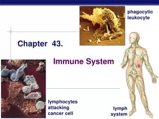
Chapter 43 The Immune System
Innate and Acquired Immunity • Innate immunity • Is present before any exposure to pathogens and is effective from the time of birth • Involves nonspecific responses to pathogens • Acquired immunity (also called adaptive immunity) • Develops only after exposure to inducing agents such as microbes, toxins, or other foreign substances • Involves a very specific response to pathogens
INNATE IMMUNITY Rapid responses to a broad range of microbes ACQUIRED IMMUNITY Slower responses to specific microbes External defenses Internal defenses Skin Phagocytic cells Humoral response (antibodies) Mucous membranes Antimicrobial proteins Secretions Inflammatory response Invading microbes (pathogens) Cell-mediated response (cytotoxic lymphocytes) Natural killer cells Summary of innate and acquired immunity
Innate Immunity • Innate immunity provides broad defenses against infection • A pathogen that successfully breaks through an animal’s external defenses soon encounters several innate cellular and chemical mechanisms that impede its attack on the body
External Defenses • Intact skin and mucous membranes • Form physical barriers that bar the entry of microorganisms and viruses • Certain cells of the mucous membranes produce mucus that traps microbes and other particles
10m First-line Respiratory Defenses • In the trachea, ciliated epithelial cells • Sweep mucus and any entrapped microbes upward, preventing the microbes from entering the lungs
Secretions • Secretions of the skin and mucous membranes • Provide an environment that is often hostile to microbes • Secretions from the skin • Give the skin a pH between 3 and 5, which is acidic enough to prevent colonization of many microbes • Also include proteins such as lysozyme, an enzyme that digests the cell walls of many bacteria
Internal Cellular and Chemical Defenses • Internal cellular defenses depend mainly on phagocytosis • Phagocytes, types of white blood cells • Ingest invading microorganisms • Initiate the inflammatory response • Macrophages, a specific type of phagocyte • Can be found migrating through the body • Can be found in various organs of the lymphatic system
Pseudopodia surround microbes. 1 Microbes Microbes are engulfed into cell. 2 MACROPHAGE Vacuole containing microbes forms. 3 Vacuole Lysosome containing enzymes Vacuole and lysosome fuse. 4 Toxic compounds and lysosomal enzymes destroy microbes. 5 Microbial debris is released by exocytosis. 6 Phagocytic Cells • Phagocytes attach to their prey via surface receptors • And engulf them, forming a vacuole that fuses with a lysosome
Interstitial fluid bathing the tissues, along with the white blood cells in it, continually enters lymphatic capillaries. 1 Lymphatic capillary Interstitial fluid Fluid inside the lymphatic capillaries, called lymph, flows through lymphatic vessels throughout the body. 2 Adenoid Tonsil Lymphatic vessels return lymph to theblood via two largeducts that drain intoveins near the shoulders. 4 Lymph nodes Blood capillary Spleen Lymphatic vessel Tissue cells Peyer’s patches (small intestine) Within lymph nodes, microbes and foreign particles present in the circulating lymph encounter macro- phages, dendritic cells, and lymphocytes, which carry out various defensive actions. 3 Appendix Lymphatic vessels Masses of lymphocytes and macrophages Lymph node • The lymphatic system plays an active role in defending the body from pathogens
Antimicrobial Proteins • Numerous proteins function in innate defense • By attacking microbes directly or by impeding their reproduction • About 30 proteins make up the complement system • Which can cause lysis of invading cells and help trigger inflammation • Interferons • Provide innate defense against viruses and help activate macrophages
Inflammatory Response • In local inflammation, histamine and other chemicals released from injured cells • Promote changes in blood vessels that allow more fluid, more phagocytes, and antimicrobial proteins to enter the tissues • 4 Classical Signs of Inflammation • Redness • Heat • Swelling • Pain
Blood clot Pin Pathogen Macrophage Blood clotting elements Chemical signals Phagocytic cells Phagocytosis Capillary Red blood cell Fluid, antimicrobial proteins, and clotting elements move from the blood to the site. Clotting begins. 1 Chemical signals released by activated macrophages and mast cells at the injury site cause nearby capillaries to widen and become more permeable. Neutrophils and macrophages phagocytose pathogens and cell debris at the site, and the tissue heals. Chemokines released by various kinds of cells attract more phagocytic cells from the blood to the injury site. 4 2 3 Major events in the local inflammatory response
Natural Killer Cells • Natural killer (NK) cells • Patrol the body and attack virus-infected body cells and cancer cells • Trigger apoptosis in the cells they attack
Invertebrate Immune Mechanisms • Many invertebrates defend themselves from infection by many of the same mechanisms in the vertebrate innate response
Acquired Immunity • Acquired immunity • Is the body’s second major kind of defense • lymphocytes provide specific defenses against infection
Antigen- binding sites Epitopes (antigenic determinants) Antibody A Antigen Antibody B Antibody C Antigens • An antigen is • Any foreign molecule that is specifically recognized by lymphocytes and elicits a response from them • A lymphocyte actually recognizes and binds to just a small, accessible portion of the antigen called an epitope
Antigen Recognition by Lymphocytes • The vertebrate body is populated by two main types of lymphocytes • B lymphocytes (B cells) and T lymphocytes (T cells) which circulate through the blood • The plasma membranes of both B cells and T cells • Have about 100,000 antigen receptor that all recognize the same epitope
Antigen- binding site Antigen- binding site Disulfide bridge V V V V Variable regions Light chain C C Constant regions C C Transmembrane region Plasma membrane Heavy chains B cell Cytoplasm of B cell (a) A B cell receptor consists of two identical heavy chains and two identical light chains linked by several disulfide bridges. B Cell Receptors for Antigens • B cell receptors bind to specific, intact antigens • Secreted antibodies are structurally similar to B cell receptors but aren’t anchored to a membrane
Antigen- Binding site Variable regions Constant regions Transmembrane region b chain Plasma membrane a chain Disulfide bridge T cell Cytoplasm of T cell (b) • A T cell receptor consists of one • chain and one b chain linked by a disulfide bridge. T Cell Receptors for Antigens and the Role of the MHC • Each T cell receptor consists of two different polypeptide chains V V C C
T cells bind to small fragments of antigens • That are bound to normal cell-surface proteins called MHC molecules • MHC molecules • Are encoded by a family of genes called the major histocompatibility complex
Infected cells produce MHC molecules • Which bind to antigen fragments and then are transported to the cell surface in a process called antigen presentation • A nearby T cell can then detect the antigen fragment displayed on the cell’s surface
Depending on their source • Peptide antigens are handled by different classes of MHC molecules
1 Infected cell A fragment of foreign protein (antigen) inside the cell associates with an MHC molecule and is transported to the cell surface. 1 Antigen fragment 2 Class I MHC molecule 2 The combination of MHC molecule and antigen is recognized by a T cell, alerting it to the infection. T cell receptor (a) Cytotoxic T cell • Class I MHC molecules, found on almost all nucleated cells of the body • Display peptide antigens to cytotoxic T cells
1 Antigen- presenting cell Microbe A fragment of foreign protein (antigen) inside the cell associates with an MHC molecule and is transported to the cell surface. 1 Antigen fragment 2 2 Class II MHC molecule The combination of MHC molecule and antigen is recognized by a T cell, alerting it to the infection. T cell receptor Helper T cell (b) • Class II MHC molecules, located mainly on dendritic cells, macrophages, and B cells • Display antigens to helper T cells
Bone marrow Lymphoid stem cell Thymus T cell B cell Blood, lymph, and lymphoid tissues (lymph nodes, spleen, and others) Lymphocyte Development • Lymphocytes arise from stem cells in the bone marrow • Newly formed lymphocytes are all alike but they later develop into B cells or T cells, depending on where they continue their maturation
Testing and Removal of Self-Reactive Lymphocytes • As B and T cells are maturing in the bone and thymus • Their antigen receptors are tested for possible self-reactivity • Lymphocytes bearing receptors for antigens already present in the body • Are destroyed by apoptosis or rendered nonfunctional
Clonal Selection of Lymphocytes • In a primary immune response • Binding of antigen to a mature lymphocyte induces the lymphocyte’s proliferation and differentiation, a process called clonal selection
Antigen molecules bind to the antigen receptors of only one of the three B cells shown. Antigen molecules B cells that differ in antigen specificity Antigen receptor The selected B cell proliferates, forming a clone of identical cells bearing receptors for the selecting antigen. Some proliferating cells develop into short-lived plasma cells that secrete antibodies specific for the antigen. Some proliferating cells develop into long-lived memory cells that can respond rapidly upon subsequent exposure to the same antigen. Antibody molecules Clone of memory cells Clone of plasma cells Clonal selection of B cells • Generates a clone of short-lived activated effector cells and a clone of long-lived memory cells
1 Secondary response to anti- gen A produces antibodies to A; primary response to anti- gen B produces antibodies to B Day 1: First exposure to antigen A 4 Primary response to antigen A produces anti- bodies to A 2 Day 28: Second exposure to antigen A; first exposure to antigen B 3 104 103 Antibody concentration (arbitrary units) 102 Antibodies to A Antibodies to B 101 100 35 28 21 42 49 56 0 14 7 Time (days) Secondary immune response • Memory cells facilitate a faster, more efficient response
Humoral and cell-mediated immunity • Acquired immunity includes two branches • The humoral immune response involves the activation and clonal selection of B cells, resulting in the production of secreted antibodies • The cell-mediated immune response involves the activation and clonal selection of cytotoxic T cells
Cell-mediated immune response Humoral immune response First exposure to antigen Antigens displayedby infected cells Antigens engulfed and displayed by dendritic cells Intact antigens Activate Activate Activate Secreted cytokines activate B cell HelperT cell CytotoxicT cell Gives rise to Gives rise to Gives rise to Active and memory helperT cells Memory cytotoxicT cells Active cytotoxicT cells MemoryB cells Plasmacells Defend against infected cells, cancer cells, and transplanted tissues Secrete antibodies that defend againstpathogens and toxins in extracellular fluid Acquired immune response
The granzymes initiate apoptosis within the target cells, leading to fragmentation of the nucleus, release of small apoptotic bodies, and eventual cell death. The released cytotoxic T cell can attack other target cells. The activated T cell releases perforin molecules, which form pores in the target cell membrane, and proteolytic enzymes (granzymes), which enter the target cell by endocytosis. A specific cytotoxic T cell binds to a class I MHC–antigen complex on a target cell via its TCR with the aid of CD8. This interaction, along with cytokines from helper T cells, leads to the activation of the cytotoxic cell. Cytotoxic T cell Released cytotoxic T cell Perforin Cancer cell Granzymes Apoptotic target cell TCR CD8 Class I MHC molecule Pore 1 3 3 2 1 2 Target cell Peptide antigen Cytotoxic T cell Figure 43.16 Activated cytotoxic T-cell • The activated cytotoxic T cell secretes proteins (perforin and granzymes) that destroy the infected target cell
Humoral Immunity ..\images and movies\anim0066-mov_humoral_im.mov
IgM (pentamer) First Ig class produced after initial exposure to antigen; then its concentration in the blood declines Promotes neutralization and agglutination of antigens; very effective in complement activation (see Figure 43.19) J chain Most abundant Ig class in blood; also present in tissue fluids IgG (monomer) Only Ig class that crosses placenta, thus conferring passive immunity on fetus Promotes opsonization, neutralization, and agglutination of antigens; less effective in complement activation than IgM (see Figure 43.19) Present in secretions such as tears, saliva, mucus, and breast milk IgA (dimer) Provides localized defense of mucous membranes by agglutination and neutralization of antigens (see Figure 43.19) J chain Secretory component Presence in breast milk confers passive immunity on nursing infant IgE (monomer) Triggers release from mast cells and basophils of histamine and other chemicals that cause allergic reactions (see Figure 43.20) IgD (monomer) Present primarily on surface of naive B cells that have not been exposed to antigens Acts as antigen receptor in antigen-stimulated proliferation and differentiation of B cells (clonal selection) Transmembrane region Antibody Classes • The five classes of immunoglobulins
Active Immunity • Active immunity • Develops naturally in response to an infection • Can also develop following immunization (vaccination) • In immunization a nonpathogenic form of a microbe or part of a microbe elicits an immune response to an immunological memory for that microbe
Passive Immunity • Passive immunity, which provides immediate, short-term protection • Is conferred naturally when IgG crosses the placenta from mother to fetus or when IgA passes from mother to infant in breast milk • Can be conferred artificially by injecting antibodies into a nonimmune person
Tissue transplantation • The immune system’s ability to distinguish self from nonself limits tissue transplantation • The immune system can attack cells from other individuals • Transplanted tissues are usually destroyed by the recipient’s immune system
Tissue and Organ Transplants • MHC molecules • Are responsible for stimulating the rejection of tissue grafts and organ transplants • The chances of successful transplantation are increased • If the donor and recipient MHC tissue types are well matched • If the recipient is given immunosuppressive drugs
Problematic Immune Responses • Exaggerated, self-directed, or diminished immune responses can cause disease • If the delicate balance of the immune system is disrupted • The effects on the individual can range from minor to fatal consequences
Allergies • Allergies are exaggerated (hypersensitive) responses to certain antigens called allergens • In localized allergies such as hay fever • IgE antibodies produced after first exposure to an allergen attach to receptors on mast cells • The next time the allergen enters the body it binds to mast cell–associated IgE molecules • The mast cells then release histamine and other mediators that cause vascular changes and typical symptoms
IgE Allergen Histamine 1 3 2 Granule Mast cell Degranulation of the cell, triggered by cross-linking of adjacent IgE molecules, releases histamine and other chemicals, leading to allergy symptoms. 1 IgE antibodies produced in response to initial exposure to an allergen bind to receptors or mast cells. 2 3 On subsequent exposure to the same allergen, IgE molecules attached to a mast cell recog- nize and bind the allergen. The allergic response
Anaphylactic shock • An acute allergic response sometimes leads to anaphylactic shock • A whole-body, life-threatening reaction that can occur within seconds of exposure to an allergen
Autoimmune Diseases • In individuals with autoimmune diseases • The immune system loses tolerance for self and turns against certain molecules of the body • Examples • Systemic lupus erythematosus • Multiple sclerosis • Insulin-dependent diabetes • Rheumatoid arthritis
Immunodeficiency Diseases • An inborn or primary immunodeficiency • Results from hereditary or congenital defects that prevent proper functioning of innate, humoral, and/or cell-mediated defenses • An acquired or secondary immunodeficiency • Results from exposure to various chemical and biological agents
Inborn (Primary) Immunodeficiencies • In severe combined immunodeficiency (SCID) • Both the humoral and cell-mediated branches of acquired immunity fail to function
Acquired (Secondary) Immunodeficiencies • Acquired immunodeficiencies range from temporary states to chronic diseases • Growing evidence shows that physical and emotional stress can harm immunity
Acquired Immunodeficiency Syndrome (AIDS) • People with AIDS are highly susceptible to opportunistic infections and cancers that take advantage of an immune system in collapse • Because AIDS arises from the loss of helper T cells both humoral and cell-mediated immune responses are impaired
1µm HIV Infection • The loss of helper T cells results from infection by the human immunodeficiency virus (HIV) Infected T-cell releasing HIV particles (gray)