Download

1 / 52
680 likes | 2k Vues
Welcome Glandular workshop 29 th May 2008. Glandular Cytology in the BD SurePath® Liquid-based Pap Test. Advanced Customer Training And Education BD Diagnostics, Diagnostic Systems – TriPath Anneke van Driel-Kulker Ph.D. Erembodegem, 29-30 May 2008. Objectives.

E N D
Welcome Glandular workshop 29th May 2008
Glandular Cytology in the BD SurePath® Liquid-based Pap Test Advanced Customer Training And Education BD Diagnostics, Diagnostic Systems – TriPath Anneke van Driel-Kulker Ph.D. Erembodegem, 29-30 May 2008
Objectives • Understand the spectrum of normal glandular cells encountered in SurePath liquid-based preparations • Identify morphologic changes seen in glandular lesions of endocervical and endometrial origin. • Review common pitfalls in glandular cytology – tubal metaplasia and lower uterine segment sampling
Cell Transfer After Sample Collection • Detach device head(s) and place into BD SurePath™vial • 100% of collected sample goes into BD SurePath™vial • Device head(s) remain in BD SurePath™vial throughout entire slide preparation process • 25% ethanol BD PrepStain™ System Product Insert — Doc 779-10002-02 Rev C BD SurePath™ Collection Product Insert — Doc. No. 779-10001-02 Rev C
“The Cell Enrichment Process” First centrifugation 2 min @ 200 rcf Second centrifugation 10 min @ 800 rcf Before Cell Enrichment After Cell Enrichment Vortex Mix and Aspirate ≈ 8 ml Layer sample solution over density gradient Photos compliments of Dr. Dugald Taylor Operator’s Manual – PrepStain slide Processor – 780-06181-00 Rev B
Slide Preparation and Staining Re-suspend cell pellet Transfer to settling chamber Stain or Prep only 01-554
Endocervical Cell Abnormalities: Atypical Glandular Cells Adenocarcinoma in-situ Endocervical Adenocarcinoma
AIS Atypical endocervical cells
AIS Adenocarcinoma in-situ
AIS Adenocarcinoma in-situ
Adenocarcinoma in-situ SurePath™ liquid-based Pap test
Adenocarcinoma in-situ • Cells occur in hyperchromatic crowded groups, strips, and rosettes with loss of honeycomb pattern • Palisading nuclei with overlap and pseudostratification common • Feathering still occurs, but may be less conspicuous • Single cells more common • Enlarged, variably sized oval or elongated nuclei – nucleoli may be present • Nuclear hyperchromasia with evenly distributed, moderate to coarse “Peppery” chromatin • Nuclear/cytoplasmic ratios increased • Mitotic figures and apoptotic bodies may be seen • Clean background • May have co-existing squamous lesion present
Endocervical adenocarcinoma Single cells, two-dimensional sheets, or three-dimensional clusters Granular or vacuolated cytoplasm Stratified and palisading nuclear arrangement Enlarged, crowded nuclei with moderate to marked anisonucleosis Fine to coarse, irregularly distributed chromatin, parachromatin clearing and nuclear membrane irregularities Presence of nucleoli and tumor diathesis May have co-existing squamous lesion present
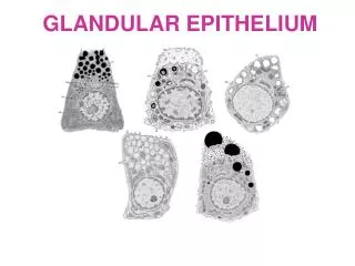
Common Pitfalls: Tubal Metaplasia and Lower Uterine Segment Sampling
Tubal Metaplasia: Crowded, honeycombed sheets and strips Nuclei are round to oval and may be enlarged, hyperchromatic, and pseudostratified Finely granular, evenly distributed chromatin Presence of cilia most helpful criterion Clean Background
Adenocarcinoma in-situ Adenocarcinoma in-situ Lower uterine segment sampling
When in doubt: • Carefully check clinical data • recent pregnancy? • patient wears IUD? • recent conisation? • Presence of stromal cells…..be aware of directly sampled endometrial cells. • Presence of cilia….tubal metaplasia.
Endocervical Lesions vs. High Grade SIL Involving Gland Spaces
High Grade SIL Involving Gland Spaces • Peripheral pallisading of nuclei, giving the appearance of glandular differentiation • Nucleoli may be present • Nuclei tend to flatten at the periphery of the cluster, creating a smooth border • Loss of central polarity and piling • Definitive glandular features are absent (pseudostratified strips, rosettes)
Endometrial Abnormalities • Atypical Endometrial Cells • Endometrial Adenocarcinoma
Atypical endometrial cells: biopsy – Grade II endometrial adenocarcinoma
Atypical endometrial cells – IUD changes Atypical endometrial cells – IUD changes
Endometrial Adenocarcinoma 3-Dimensional groups with scalloped borders, papillary configurations, and single cells Variation in nuclear size; nuclei become larger with increasing tumor grade Nucleoli may be small to prominent and become larger with increasing tumor grade Nuclear hyperchromasia, irregular chromatin distribution and clearing Intracytoplasmic neutrophils and vacuoles common Cleaner background – tumor diathesis may be less prominent
Importance of clinical data • 90% of endometrium cancers is post menopausal and cause bleeding. • Almost all endometroid cancers produce estrogen and therefore squamous cells are well matured. • Endometrium cells in smears from highly differentiated adenocarcinoma of the endometrium can be cytomorphologically normal. • IUD can cause severe cytomorphological changes to the endometrium cells: irregularity and hyperchromasia of nuclei, macronucleoli.
Rare !! • Glandular abnormalities constitute <2% of all abnormalities in cervical cytology • Glandular lesions are the most interesting AND the most difficult of all cervical cytology • Experience with LBC in combination with sampling device is needed to correctly interpret atypical glandular cells.