Download
1 / 25
250 likes | 491 Vues
Case JHC. Juan G. Santiago, MD Ophthalmology Department University of Puerto Rico. Chief Complaint. “ Veo borroso por el ojo izquierdo hace 4 semanas ”. Present History.

E N D
Case JHC Juan G. Santiago, MD Ophthalmology Department University of Puerto Rico
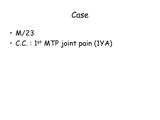
Chief Complaint • “Veoborrosopor el ojoizquierdohace 4 semanas”
Present History • JHC is a 58 y/o male with history of HTN, heart disease in his usual state of health that complaints of left eye visual loss since 4 weeks ago. Patient refers a gray cloud in front of his eye, that does not move. Denies improvement or worsening of vision since event. Patient refers (-) eye pain, (-) photophobia, (-) flashlights, (-) diplopia, (-) secretions, (-) redness, (-) pain with eye movements, (-) headaches, (-) jaw claudication, (-) extremity weakness, (-) trauma, or (-) recent illness.
History • Eye history: None • Surgeries: None • Childhood: None • Systemic history: HTN, Heart Disease • Family history: BA, DM, HTN
Exam BP: 170/92
Differential Diagnosis • Nonarteritic Anterior ION • Pseudo Foster-Kennedy Syndrome • Compressive Optic Neuropathy • True Foster-Kennedy Syndrome • Malignant Hypertension • Inflammatory Optic Neuropathy • Infectious Optic Neuropathy • Arteritic Anterior ION
Pseudo Foster Kennedy Syndrome Bilateral sequential infarction of optic nerve Sudden and painless visual loss Unilateral disc edema + Optic atrophy Flame shaped hemorrhages Usually > 50 yrs Nonarteritic Anterior ION
Nonarteritic Anterior ION • DM / HTN • Contralateral small C/D ratio • ↓ Color vision • VF loss (altitudinal) • No associated symptoms • Normal ESR and C-RP • Treatment: NONE
True Foster Kennedy Syndrome Large mass lesion compressing the atrophic optic nerve and elevating intracranial pressure, to cause disc swelling in the other nerve Slow progressive visual loss ↓ Color vision Visual field defect Compressive Optic Neuropathy
Etiologies ON tumor Pituitary tumor or apoplexy Diagnosis Orbit CT/MRI Compressive Optic Neuropathy
Malignant Hypertension • Very high blood pressure (>200/130) with papilledema • Accompanied by • Heart failure • Kidney failure • Hypertensive encephalopathy
Flame-shaped hemorrhages Cotton-wool spots Exudative edema Vessel sheathing Vessel tortuosity Malignant Hypertension
Inflammatory and Infectious Optic Neuropathies • History is atypical • Atypical clinical course • Ongoing pain • Lack of visual recovery • Recurrence on steroid taper • Associated signs of intraocular infection • Optic disc appearance is not typical • Imaging is not typical • Patient has other systemic illness
Sarcoidosis Infectious Viral Syphilis Lyme disease Bartonella TB Autoimmune Wegener’s SLE Devic’s syndrome Inflammatory and InfectiousOptic Neuropathies
Arteritic Anterior ION • Infarction of optic nerve 2ry to temporal arteritis • Acute painful visual loss • Female > male (2:1) • Aged >65 yrs • ↓ Color vision • VF loss • Unilateral OD edema • Amaurosis fugax or diplopia
Arteritic Anterior ION Associated symptoms • Scalp tenderness • Jaw claudication • Fever • Malaise • Anorexia • Weight loss • Anemia • Headache • Tender temporal artery Diagnosis: • ↑ ESR • ↑ C-RP • ↓ HCT / ↑ PLT • Temporal artery biopsy • Inflammation in artery wall with disruption of internal elastica lamina
Our patient… • Head and Orbit CT • Preliminar report: Enlarged left optic nerve??? • Labs • CBC WNL • RPR Non-Rx • ANA Screen Neg • HIV Non-Rx • Chest X-rays • Normal • No hilaradenopathies • CSF WNL • ESR 8 • CRP 0.252 • Vit B12 446
Our patient… • Head and Orbit CT • WNL • No evidence of enlarged optic nerves, no masses • Brain MRI • WNL
Our diagnosis… • Pseudo Foster Kennedy Syndrome • Bilateral Sequential NAAION