Download
1 / 60
610 likes | 766 Vues
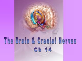
Chapter 12 & 13 The Brain and Cranial Nerves. Central Nervous System (CNS). CNS: Composed of the brain and spinal cord Brain: Control center for many of body’s functions Is composed of wrinkled, pinkish gray tissue Consists of Cerebrum Cerebellum Diencephalon Brain stem.

E N D
Chapter 12 & 13 The Brain and Cranial Nerves
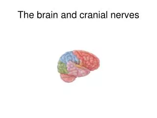
Central Nervous System (CNS) • CNS: Composed of the brain and spinal cord • Brain: Control center for many of body’s functions • Is composed of wrinkled, pinkish gray tissue • Consists of • Cerebrum • Cerebellum • Diencephalon • Brain stem
Embryonic Development • During the first 26 days of development: • Ectoderm thickens forming the neural plate • The neural plate invaginates, forming the neural groove • The neural groove fuses dorsally and forms the neural tube
Primary Brain Vesicles • The anterior end of the neural tube expands and constricts to form the three primary brain vesicles • Prosencephalon – the forebrain • Mesencephalon – the midbrain • Rhombencephalon – hindbrain
Secondary Brain Vesicles • In week 5 of embryonic development, secondary brain vesicles form • Telencephalon and diencephalon arise from the forebrain • Mesencephalon remains undivided • Metencephalon and myelencephalon arise from the hindbrain
Adult Brain Structures • Secondary brain vesicles develop into: • Telencephalon – cerebrum • Diencephalon – thalamus, hypothalamus, and epithalamus • Mesencephalon – brain stem: midbrain • Metencephalon –Cerebellum and brain stem: pons • Myelencephalon – brain stem: medulla oblongata
Largest portion of brain Composed of right and left hemispheres Contain numerous folds, increase surface area - Gyri Shallow grooves or valleysbetween gyri - Sulci Contain deeper grooves - Fissures Cerebrum
Sulci and Fissures: Longitudinal fissure: separates the two hemispheres Lateral fissure: separates temporal lobe from frontal and parietal lobes Central sulcus: separates frontal and parietal lobes Cerebrum
Each Cerebral hemisphere is divided into different lobes: Frontal lobe: Imp. in voluntary motor function, motivation, aggression, sense of smell, mood Parietal lobe: Center for reception and evaluation of sensory information except smell, hearing, and vision Cerebrum
Occipital lobe: reception and integration of visual input Temporal lobe: reception and evaluation for smell and hearing; memory, abstract thought, judgment Insula:Insula is present within temporal lobe – 5th lobe Play important role in emotions, regulate body`s homeostasis Cerebrum
Cerebral cortex: outer surface, consists of gray matter Contains no. of neurons such as fusiform cells, stellate cells, pyramidal cells Gray matter deep inside the brain – Basal nuclei Cerebral Medulla:White matter between the cortex & nuclei Consists of deep myelinated fibers and their tracts It is responsible for communication between: Cerebral cortex & lower CNS center & areas of the cerebrum Cerebrum
Three types of fibers in cerebral white matter: Association fibers: connect area of cerebral cortex within the same hemisphere Commissural fibers: connect one hemisphere to the other hemisphere – Corpus callosum Projection fibers: connect cerebrum to other parts of the brain and spinal cord Cerebrum
Masses of gray matter found deep within the white matter of cerebrum, diencephalon & midbrain– basal nuclei Nuclei are involved in the control of motor functions Nuclei in cerebrum are called Corpus Striatum and composed of: Caudate nucleus Lentiform nucleus Lentiform nucleus composed of: - putamen - Globus pallidus Basal Nuclei
Basal Nuclei are largest nuclei of brain and occupy largest part of cerebrum Sub thalamic nucleus : Present in diencephalon Substantia nigra : Present in midbrain Both these nuclei function with cerebrum basal nuclei to control movement Basal Nuclei
Part of cerebrum and diencephalon Plays a role in Emotions andbasic survival functions such as memory, reproduction, nutrition Limbic system consists of : Cingulate gyrus & Para hippocampal gyrus of Cerebrum Various nuclei of the thalamus Part of the basal nuclei, hypothalamus, olfactory cortex Limbic System
Located between brainstem and cerebrum Consists of – Thalamus Sub thalamus Hypothalamus Epithalamus Diencephalon
80% of diencephalon Two large gray masses connected by interthalamic adhesion or intermediate mass Many nuclei Sensory relay center of the brain All sensory neurons except olfactory neurons first synapse in thalamus Then thalamus directs information to proper cortical region Regulate emotions, moods, motor activities, learning, and memory Thalamus
Small area inferior to thalamus Contains several ascending and descending nerve tracts Contains sub thalamic nuclei, parts of red nuclei and substantia nigra Involved in controlling motor function Sub thalamus
Most inferior portion of diencephalon, located below the thalamus Contains several nuclei and tracts Mammillary bodies Small, paired nuclei bulging anteriorly from the hypothalamus Relay station for olfactory pathway Response to odor Infundibulum –stalk of the hypothalamus; connects hypothalamus to posterior pituitary gland Hypothalamus controls endocrine system Regulates Pituitary gland Hypothalamus
Initiates physical expression of emotions (linked to the limbic system) Regulates thirst, food intake, body temp., sexual behavior, pleasant and painful feelings, pleasure, fear, rage, sleep-wake cycles Regulates autonomic centers in the brain stem controlling B.P., digestive rate, respiration rate Hypothalamic Function
Small area superior and posterior to thalamus Consists of Pineal body and Habenular nucleus Pineal gland – Secretes melatonin Melatonin helps regulate sleep-wake cycles Pineal gland may play a role in controlling the onset of puberty Habenular nucleus: Involved in emotional and visceral responses to odors Epithalamus
Consists of three regions – Midbrain Pons Medulla Oblongata Connects spinal cord to the remainder of brain Associated with 10 of the 12 pairs of cranial nerves Damage to brainstem causes death Because many reflexes essential for survival are integrated in brainstem Brain Stem
Located between the diencephalon and the pons Midbrain structures include: Cerebral peduncles – two bulging structures, consists of descending tracts Which carry motor information from cerebrum to Brain Stem & Spinal cord Cerebral aqueduct – narrow canal that connects the third and fourth ventricle Various nuclei Brain Stem: Midbrain
Nuclei that control cranial nerves III (oculomotor), IV (trochlear) and V (trigeminal) Corpora quadrigemina – four dome shaped nuclei on the dorsal midbrain - Superior colliculi – 2 Superior nuclei, visual reflex centers, coordinate head and eye movements - Inferior colliculi – 2 Inferior nuclei, auditory relay centers, Act in reflexive responses to sound Brain Stem: Midbrain Nuclei
Substantia nigra – Interconnected with basal nuclei of cerebrum, involve in coordinating movements Contains melanin pigment Precursor of dopamine neurotransmitter Degeneration of dopamine-releasing neurons – Parkinson`s disease Red nucleus – Reddish due to rich blood supply and iron content Aid in unconscious regulation and coordination of motor activities Brain Stem: Midbrain Nuclei
Superior to the medulla oblongata Contains ascending & descending tracts Many Nuclei Pontine nuclei: anterior portion of pons, relay information between cerebrum and cerebellum Nuclei for cranial nerves V-IX: located in posterior pons Pons nuclei are the part of - Sleep center - Help medulla oblongata to control respiratory movements Brain Stem: Pons
Most inferior part of brain & continuous with SC Has both ascending and descending nerve tracts Many medullary nuclei functions as: Regulate heart rate, blood vessel diameter, respiration, swallowing, vomiting, hiccupping, coughing, and sneezing Brain Stem: Medulla Oblongata
Pyramids: 2 anterior bulges Contains descending nerve tracts on the anterior surface. Inferiorly fibers decussate (cross over); thus each half of the brain controls the opposite half of the body Olives: 2 rounded structure; protrude from anterior surface. Nuclei within help regulate balance, coordination, modulation of sound from inner ear Nuclei of cranial nerves V, IX-XII Brain Stem: Medulla Oblongata
Attached to brainstem posterior to pons Three paired fiber tracts that connect the cerebellum to the brain stem Superior peduncles connect the cerebellum to the midbrain Middle peduncles connect the pons to the cerebellum Inferior peduncles connect the medulla oblongata to the cerebellum Cerebellum
Two bilaterally symmetrical hemispheres connected medially by the vermis Folia –Ridges of Cerebellar cortex Arbor vitae – White matter with a tree- like appearance Cerebral cortex contains several types of neurons including: stellate, basket, granule, Golgi cells, Purkinje cells – largest cell in CNS Cerebellar cortex contains more than 1012 neurons, more neurons than entire Cerebral cortex Cerebellum
Cerebellum consists of 3 parts: Flocculonodular lobe: a small inferior part Vermis: narrow central worm like structure Two lateral hemispheres Cerebellum
Flocculonodular lobe: Control balance and eye movements Vermis and medial portion of hemispheres: Control posture, locomotion, fine motor coordination Lateral hemispheres, major portion: works with cerebrum to plan, practice, learn complex movements Cerebellar Functions
Three connective tissue membranes lie external to the CNS – dura mater, arachnoid mater, and pia mater Functions of the meninges Cover and protect the CNS Protect blood vessels and enclose venous sinuses Contain cerebrospinal fluid (CSF) Form partitions within the skull Meninges
Outermost, strong meninx composed of two fibrous connective tissue layers The two layers separate in certain areas and form Dural venous sinuses All veins drain blood from brain empty into Dural venous sinuses Dura mater extends inward in some areas forming septa to anchor the brain to the skull Dura Mater
Three Dural septa extend into major brain fissures and limit excessive movement of the brain Falx cerebri – fold that dips into the longitudinal fissure Falx cerebelli – runs along the vermis of the cerebellum Tentorium cerebelli – horizontal Dural fold extends into the transverse fissure Dura Mater
The middle meninx, which forms a loose brain covering It is separated from the dura mater by the subdural space Arachnoid Mater
Pia mater: thin, delicate C.T. membrane closely attached to the surface of the brain Subarachnoid space: Present between pia mater and subarachnoid mater Contains blood vessels, and cerebrospinal fluid Pia Mater
Ventricles • Brain ventricles arise from expansions of the lumen of embryonic neural tube • Interconnected chambers within brain • Ventricular chambers are filled with CSF and lined by ependymal cells • Lateral ventricles: large cavities within cerebral hemispheres • Third ventricle: within diencephalon • Interventricular foramina join lateral ventricles with third ventricle
Ventricles • Fourth ventricle: • Dorsal to pons and medulla • 4th ventricle connected to 3rd ventricle by the cerebral aqueduct • Opens into central canal of spinal cord • And also connected to the subarachnoid space by two lateral & one medial apertures
Similar to serum, but most protein removed Flows around and in the brain and spinal cord Protective cushion around CNS Prevents the brain from crushing under its own weight Protects the CNS from blows and other trauma Nourishes the brain Cerebrospinal Fluid (CSF)
99% water Also sugar (glucose), chlorides, few proteins, ions, vitamin C Total volume of 150 ml (replaced every 8 hrs.) 500 ml formed daily Formed by choroid plexuses in the brain ventricles Choroid plexuses are composed of ependymal cells, their support tissue, and associated blood vessels Cerebrospinal Fluid (CSF)
Endothelial cells (lining all capillaries) have tight junctions between them Astrocytes have foot processes that influence capillary permeability Basement membrane of endothelium Excludes many potentially harmful substances Ineffective against some substances Fats and fat soluble molecules Respiratory gases Alcohol Nicotine Anesthesia Blood-Brain Barrier
Brain Requires a tremendous amount of blood Receives 15-20% of blood pumped by heart Interruption can cause unconsciousness and irreversible brain damage High metabolic rate; dependent upon constant supply of oxygen and glucose Receives blood through arteries: internal carotids and vertebral arteries. The vertebral arteries join to form the basilar artery. Carotids plus basilar form the cerebral arterial circle (Circle of Willis). Blood Supply to the Brain
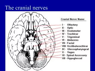
Cranial Nerves Olfactory nerve (I) optic chiasma Optic nerve (II) Oculomotor nerve (III) Trochlear nerve (IV) Abducens nerve (VI) Trigeminal nerve (V) Vestibulocochlear nerve (VIII) Facial nerve (VII) Vagus nerve (X) Glossopharyngeal nerve (IX) Hypoglossal nerve (XII) Accessory nerve (XI)
12 pair numbered by their location (from anterior to posterior) C.N. (I) and (II) attach to the cerebrum/diencephalon C.N. (III) through (XII) attach to the brainstem Almost all of the cranial nerves serve the head and neck C.N. (X), Vagus, extends into the abdominal cavity Cranial nerves can have sensory, motor or parasympathetic neuron fibers (or all of these) C.N. (III), (VII), (IX), and (X) contain parasympathetic fibers OldOpieOccasionally TriesTrigonometry AndFeelsVery Gloomy,Vague,AndHypoactive Cranial Nerves
Cranial Nerve I : Olfactory • Sensory only • Carry smell sensations from nasal cavity • Originate in nasal cavity • Pass through the cribriform plate of ethmoid bone to olfactory bulb • Olfactory nerves olfactory bulb olfactory tract inferior frontal and medial temporal lobes
Sensory only Originate from the retina of the eye Optic nerves optic chiasma optic tracts lateral geniculate bodies of thalamus optic radiations visual cortex in occipital lobe Function: carry afferent impulses for vision Cranial Nerve II : Optic
Motor, sensory and parasympathetic fibers Fibers extend from the ventral midbrain, pass through the superior orbital fissure, and go to the extrinsic eye muscles Functions in raising the eyelid, directing the eyeball, constricting the iris, and controlling lens shape Cranial Nerve III : Oculomotor
Motor and sensory Primarily a motor nerve that directs the eyeball Fibers emerge from the dorsal midbrain and enter the orbits via the superior orbital fissures; innervate the superior oblique muscle Cranial Nerve IV: Trochlear