Download
1 / 48
570 likes | 1.3k Vues
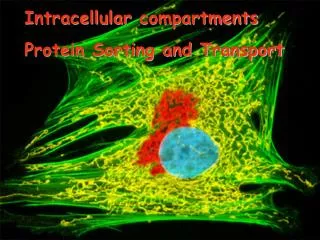
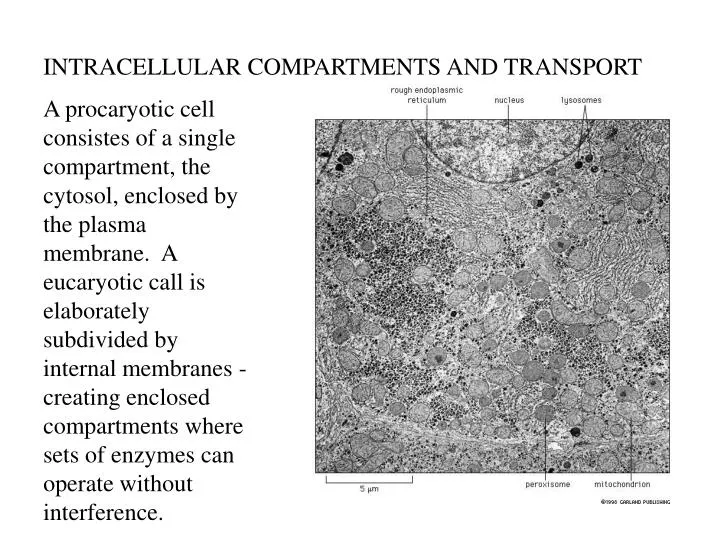
INTRACELLULAR COMPARTMENTS AND TRANSPORT. A procaryotic cell consistes of a single compartment, the cytosol, enclosed by the plasma membrane. A eucaryotic call is elaborately subdivided by internal membranes - creating enclosed compartments where sets of enzymes can operate without interference.

E N D
INTRACELLULAR COMPARTMENTS AND TRANSPORT A procaryotic cell consistes of a single compartment, the cytosol, enclosed by the plasma membrane. A eucaryotic call is elaborately subdivided by internal membranes - creating enclosed compartments where sets of enzymes can operate without interference.
Many organelles are held in place by attachment to the cytoskeleton, especially microtubules. • Cytoskeletal filaments provide tracks for moving organelles around, for directing the traffic of vesicles between them • These movements are driven by motor proteins that use ATP • Total amount of membrane in eucaryotic cells is enormous, plasma membrane is only a minor membrane in most.
Hypothesis for evolution of the nuclear membranes and the ER.
Eucaryotic cells carry out thousands of chemical reactions, many of which need to be separated - glucose breakdown/glucose synthesis. • Strategies for segregating and organizing their chemical reactions include • aggregating the different enzymes required to catalyze a particular sequence of reactions into a single large multiprotein complex - ribosomes • confining different metabolic processes and the proteins required to carry them out within different membrane-bounded compartments - compartmentalization
Table 1 Know all discussed on page 449 Each compartment contains a unique set of proteins, which have to be transferred selectively from the cytosol, where they are made, to the compartment in which they are used - protein sorting. Protein sorting depends on signals built into the amino acid sequence of the proteins.
PROTEIN SORTING Membrane-bound organelles must be enlarged for cell division and maintained during the life of the cell. Lipids and proteins must be sythesized and directed appropriately Nuclear envelope, ER, and Golgi break up into small vesicles which later coalase in the daughter cells
Synthesis of most proteins begins on ribosomes in the cytosol. Few exceptions include some mitochondrial and chloroplast proteins synthesized on ribosomes in these organelles. Each protein contains a sorting signal which directs the protein to the organelle in which it is required. Lacking a sorting signal, proteins remain in the cytosol. Membrane translocators
Signal sequences are both necessary and sufficient to direct a protein to a particular organelle. Each protein contains a sorting signal which directs the protein to the organelle in which it is required. Lacking a sorting signal, proteins remain in the cytosol. Signal sequences specifying the same destination can vary greatly even though they have the same function.
Proteins enter the nucleus through nuclear pores. The composition of the outer nuclear membrane closely resembeles the ER, which is continuous. The inner membrane contains proteins that act as binding sites for chromosomes and for the nuclear lamina The nuclear envelope contains many nuclear pores which form gates through which molecules enter or leave the nucleus.
Nuclear transport through the pores is a final quality control step rRNA, along with completed ribosomal subunits are exported. mRNA is exported only upon completion.
A nuclear pore is a large elaborate structure. Water-soluble molecules can pass freely but larger molecules and macromolecular complexes cannot pass unless they carry an appropriate.sorting signal - nuclear localization signal.
Protein fibrils protrude from both sides of the complex; on the nuclear side they converge to form a cagelike structure.
Nuclear import receptors (cytosol) bind to the nuclear localization signal on the protein being transferred, bind the fibrils, and help direct it to the pore. The nuclear protein is actively transported into the nucleus using GTP hydrolysis and the nuclear import receptors return to the cytoplasm Nuclear pores transport proteins and ribosomal subunits in their fully folded conformation
Most mitochondrial and chloroplast proteins are encoded by nuclear genes and imported from the cytosol. Proteins unfold to enter mitochondria and chloroplasts. The protein is translocated simultaneously across both the inner and outer membranes at specialized sites where the two membranes are in contact with each other Chaperone proteins help pull proteins across and help it refold
In many cells the ER is the most extensive membrane system in the cell. The ER serves as an entry point for proteins destined for other organelles, as well as the ER. Once inside the ER, proteins will not reenter the cytosol. - they are ferried by transport vesicles to various organelles. Two kinds of proteins are transferrred from the cytosol to the ER: 1. Water-soluble proteins are completely translocated into the ER lumen. 2. Prospective transmembrane proteins are only partly translocated and become embedded in the ER membrane in the proper orientation
Water soluble proteins that do not remain in the ER will be secreted or will be transported into the lumen of an organelle. Transmembrane proteins that do not remain in the ER membrane are transported into the membrane of another organelle or the plasma membrane.
SRP (signal-recognition particle) binds the ER signal sequence on the mRNA. This slows protein synthesis. Once SRP bind its receptor in the ER membrane and docks with the translocation channel, it is released and protein synthesis resumes. Amino terminus
The signal sequence opens the translocation channel. The ribosome remains attached while the rest of the protein is threaded through the membrane. Signal peptidase cleaves the signal sequence.
Start and Stop signals determine the arrangement of a transmembrane protein in the lipid bilayer.
The integration of a double pass transmembrane protein. Complex multipass proteins contain further pairs of stop and start sequences.
Vesicular transport between membrane-bounded compartments is highly organized. Secretory pathway leads from biosynthesis of lipids and proteins on the ER membrane: Each transport vesicle must take with it only the proteins and lipids appropriate to its destination and must fuse only with the appropriate target membrane
Vesicle budding is driven by the assembly of a protein coat on their cytosolic surface - coated vesicles. After budding is complete, the coat is lost. There are several kinds of coated vesicles, each with distinctive protein coats. These are thought to serve at least two functions 1. Shapes the membrane into a vessicle 2. Helps to capture the appropriate molecules to be transported. The best described is the clathrin-coated vesicle.
Clathrin-coated pits and vesicles budding from the inner surface of the plasma membrane of cultured skin cells
Clathrin-coated vesicles bud from the golgi on the outward secretory pathway and from the plasma membrane on the inward endocytic pathway. Clathrin molecules assemble on the cytosolic surface of the membrane - this starts the shaping of the membrane into a vesicle. A small GTP-binding protein - dynamin - forms a ring around the neck. GTP is hydrolized, the ring constricts, pinching off the neck and releasing the vesicle.
1. How does the transport vesicle select the appropriate molecules to transport? Excluding all other? In the clathrin system, adaptins bind the coat and the vesicle membrane. These help select the molecules for transport because the adaptins bind the membrane via specific cargo receptors. Molecules to be transported carry specific transport signals that bind to specific cargo receptors. GTP --> GDP There are other classes of coated vesicles - see Table 4
2. How do the vesicles get to the appropriate destination? How do lysosomal enzymes get to the lysosome and not to the mitochondria? If the distance is short (ER to Golgi) diffusion will work. However, if the distance is longer, vesicles are actively transported by motor proteins that move along cytoskeletal fibers. The impressive specificity of vesicular transport suggests that all types of transport vesicles have surface molecular markers that identify the vesicle according to its origin and cargo. These must be recognized by complementary receptors on the target membrane.
This recognition is thought to involve a family of proteins - SNARES. Each organelle is believed to carry a unique SNARE V is for vesicle, t is for target
Docking occurs when v- and t-SNAREs bind. However, docking does not ensure fusion. Fusion requires a much closer approach, probably catalyzed by specialized proteins. A few of the cytosolic proteins required for fusion have been identified.
Secretory pathways- exocytosis Most proteins are covalently modified in the ER. 1. Disulfide bonds are formed by the oxidation of pairs of cysteine side chains catalyzed by an enzyme in the ER lumen. Disulfide bonds help stabilize the structure of proteins encountering changes in pH.
Disulfide bonds do not form in the cytosol. Remember, they are important in the extracellular matrix secreted by cells.
Most proteins are covalently modified in the ER . 2. Glycosylation - covalent attachment of short oligosaccharide side chains - results in glycoproteins. Glycosylation is carried out by enzymes in the lumin of the ER. These oligosaccharides have various functions:1. Protects proteins from degradation 2. Retain the protein in the ER until properly folded 3. Help guide it to the appropriate organelle by serving as a transport signal 4. On the cell surface they form the glycocalyx 5. Function as recognition molecules between different cells.
5. Function as recognition molecules between different cells.
A preformed branched oligosaccharide, containing 14 sugars is attached to all proteins that carry the appropriate site for glycosylation. This oligosaccharide is originally attached to a lipid – dolichol – then transferred to the amino group of an asparagine side chain. A simple sequence of 3 amino acids defines this signal. Theseare called N-linked.
Glycoproteins are remarkably diverse. These core oligosaccharide chains are modified in the ER and Golgi.
Exit from the ER is controlled to ensure protein quality. Proteins that function in the ER are retained there or returned from the Golgi by an ER retention signal which is recognized by a membrane-bound receptor protein in the ER and Golgi. All other proteins move in transport vesicles to the Golgi if they are folded and assembled properly. Chaperone proteins in the ER hold proteins until they fold and assemble properly, or are degraded. Any protein, even a functional protein, that can not fold properly will be degraded. Ex. cystic fibrosis – mutant plasma membrane transport protein
Golgi apparatus: Ordered progession through the Golgi. Ex. Many proteins have complex oligosaccharide chains created by a highly ordered process in which sugars are added or removed in a determined sequence performed by a series of enzymes positioned in the Golgi stacks in the proper order.
Secretory proteins are released from the cell by exocytosis in either a constitutive or a regulated manner. Extracellular matrix, hormones, etc hormones, mucus, digestive enzymes, etc
In secretory cells like beta cells of the pancreas, large quantities of a particular molecule, like insulin, are stored at high concentrations (aggregated) in vesicles near the plasma membrane. These cells can then respond to a signal by releasing large quantities quickly. The membrane of the vesicle becomes part of the plasma membrane. Endocytosis returns lipids and membrane proteins to the Golgi.
Endocytic pathways: Eucaryotic cells are constantly taking up liquid and large/small molecules by endocytosis. Ultimately to lysosomes for digestion. Metabolites generated are transferred directly out of the lysosome into the cytosol. Pinocytosis = ingestion of fluid and molecules via small vesicles (by all cells) Receptor-mediated endocytosis = uptake of specific molecules by any cell with the receptor for this specific molecule Phagocytosis = ingestion of large particles, such as microbes and cell debris via large vesicles called phagosomes. (by phagocytic cells)
Phagocytes are an important part of our immune system. Macrophages are found in large numbers in all connective tissue. The are on the spot when microbes break through our barriors, into our tissues. Phagocytes of our immune system also express various receptors in their plasma membrane which bind to pathogens or antibodies which are bound to pathogens. These receptors help phagocytes ingest more efficiently - receptor-mediated endocytosis.
psuedopods – fine sheets of membrane that the macrophage is extending to engulf the red blood cells in the spleen. In protozoa, phagocytosis is a form of feeding. Phagosomes fuse with lysosomes, forming phagolysosomes. Here the food is digested.
Pinocytosis is a continuous process in eucaryotic cells, therefore lipids subtracted from the membrane and fluid taken into the cell must be balanced since total surface area and volume is maintained. • Pinocytosis is mainly carried out by the clathrin-coated pits and vesicles. • Pinocytosis is indiscriminate unless it encorporates receptor-mediate endocytosis.
Receptor-mediated pinocytosis via clathrin-coated vesicles is an efficient pathway for uptake of specific marcomolecules from the extracellular fluid. *defective LDL receptor – cholesterol accumulates in the blood atherosclerosis cholesterol is insoluble and is transported in the blood via low-density lipoproteins (LDL) This method of entering a cell is used by many pathogens including HIV.
Lysosomes are membranous sacs of hydrolytic enzymes that digest both extracellular materials and worn out organelles. These enzymes are active at acidic pH. Therefore, even if these enzymes escape into the cytosol, they are not active at neutral pH. transport proteins to pump breakdown products into the cytosol for use by the cell in synthesis etc. ATP driven H+ pump