Download
1 / 41
580 likes | 1.6k Vues
Intracellular Vesicular Transport. Yasir Waheed. NUST Center of Virology & Immunolgy National University of Sciences &Technology. Central Dogma of Molecular Biology. DNA. Transcription. Transcription. Translation. mRNA. Translation. Protein.

E N D
Intracellular Vesicular Transport Yasir Waheed NUST Center of Virology & Immunolgy National University of Sciences &Technology
Central Dogma of Molecular Biology DNA Transcription Transcription Translation mRNA Translation Protein
Proteins carry signal sequence/patch for their transport form cytoplasm to ER and from ER to Golgi and other organelles
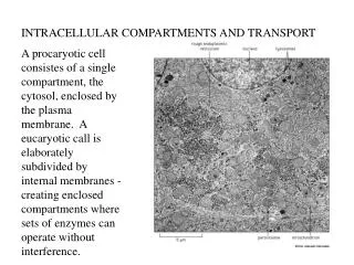
Every cell must eat, and it must communicate with the world around it • In a procaryotic cell, all the eating and communicating takes place across the plasma membrane • The cell secretes digestive enzymes, for example, across the plasma membrane to the cell exterior. It then transports the small metabolites generated by digestion in the extracellular space across the same membrane into the cytosol • Eucaryotic cells, by contrast, have evolved an elaborate internal membrane system that allows them to take up macromolecules by the process of endocytosis and deliver them to digestive enzymes stored in lysosomes inside the cell
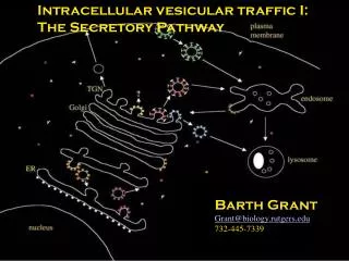
Intracellular Vesicular Transport • Intracellular Secretory pathway leads outward from ER towards the Golgi Apparatus and Cell Surface ,with a side route leading to lysosomes.
Transport Vesicle bud off from one compartment and fuse with another In doing so they carry material as cargo from the lumen and membrane of donor compartment to the lumen and membrane of the target compartment
Types of Coated Vesicles • There are three well characterize type coated vesicles, differ in their coat proteins which are • Clathrin Coated Vesicles • COP 1 Coated Vesicles • COP 2 Coated Vesicles • Each type is used for different transport step in the cell
Clathrin Coated Vesicles • The major component of Clathrin Coated vesilces is Clathrin itself. • Each Clathrin subunit consists of 3 large and 3 small Polypeptide chains that togethered form a three legged structure called Triskelion.
Second Major protein is Adaptin,which is required both to bind the Clathrin coat to the membrane and to trap various transmembrane protein including transmembrane receptors that capture soluble cargo molecules inside the vesilce so called cargo vesicles
Dynamin regulated the pinching off from the membrane. In Pinching off process. The 2 leaflets of membrane are brought into close proximity and fuse, sealing off the forming vesicle.
Once vesicle is released from the membrane the Clathrin Coat is rapidly lost A chaperone protein hsp 70 family function as uncoating ATPases, using the energy of ATP hydrolysis to peel off the coat. The assembly of coat is thought to introduce curvature into the membrane which leads in turns to the formation of the uniformly sized coat bud.
COP 1 Coated Vesilces: Bud from Golgi Apparatus COP 2 Coated Vesilces: Bud from ER
COP 2 Coated Vesicles • Control by GTPase , Active when GTP is present and inactive when GDP is present
Sar1 is coat recruitment GTPase , Active when GTP is present and inactive when GDP is present. Sar1 insert tail into the membrane of ER.
SNARE Proteins • To ensure that membrane traffic proceed in an orderly way,transport vesilce must be highly selective in recognizing the correct target membrane with which to fuse. • Vesicle encounter many membranes before finding the correct one. • Vesicle displays surface markers called Snares.
Recognition involve two classes of proteins Snares V-snare and t-snare V-snare binds with specific T-snares and lock two membranes together Targeting GTPase called Rabs Rab facilitate and regulate rate of docking.
Transport from the ER through the Golgi Appartus. COP II coated Vesilces carry cargo from ER to Golgi
Only proteins that properly folded and assemble can leave the ER. Chaperon BiP bind with the malfolded protein and degrade it.
Retrieval pathway to ER using sorting signal Proteins having the KDEL (Lys-Asp-Gly-Leu) sequence return back to ER packed in COP1 coated vesicles.
Golgi Apparatus consists of an Orderd series of compartments. • Gogli stacks has two distinct faces • Cis face (entry face) .Forming Cis Golgi Network • Trans Face (exit face). Forming Tans Golgi Network
Oligosaccharide chains are Processed in Golgi Apparatus • Two broad classes of Oligosaccharides are produced in ER which are processed in Golgi. • Complex Oligosaccharides. • High mannose oligosaccharides.
The most common modifications in glycoproteins occurs through N- or O- glycosidic bonds O-linkage: Ser, Thr (GalNAc) N-linkage: Asn (GlcNAc)
Transport from Trans Golgi Network to Lysosomes. • Lysosomes are membrane encolsed compartment use for intracellular digestion of macromolecules. • They contain about 40 types of hydrolytic enzymes including nucleases, glycosidases, lipases, phosphatases and sulfatases. All are acid hydrolases. • Require pH 5 for their activity. • If they leak from the lysosomes they • Will not cause harm at cytoplasmic pH7.2
Multiple pathways deliver materials to Lysosomes • Endocytosis: Macromolecules taken in, form early endosomes, some material taken up by the cell, while other deliver to late endosomes (pH 6) and then sent to Lysosomes(pH 5) for degradation. • Autophagy: Mitochondrial life time is about 10 days, then taken up by the lysosomes and degraded. • Phagocytosis: Bacteria are taken in by phagosome and then sent to lysosomes
Mannose 6-Phosphate recognizes Lysosomal Proteins in the Trans Golgi Network • By this mechanism the membrane proteins of lysosomes and lysosomal Hydrolases are sent to Lysosomes. • They carry M6P at their surface which are recognize by the receptors, packed and sent to lysosomes. • The M6P receptor carry KDEL sequence and come back to ER.
SUMMARY Trafficking : ER to Golgi to Lysosome Proteins are synthesize on ribosomes and sent to ER where protein folding and Glycosylation takes place. Protein is then sent to Golgi for glycosylation trimming and packing and sent to various locations like plasmamembrane, cell exterior , lysosomes , other organelles.