Download
1 / 38
400 likes | 547 Vues
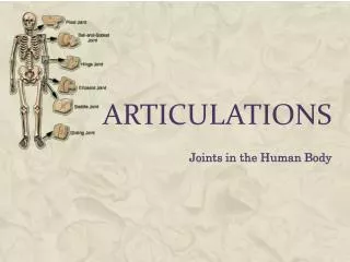
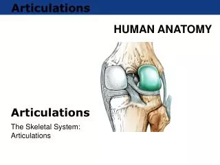
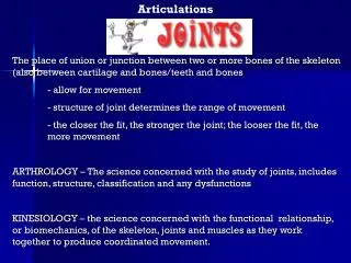
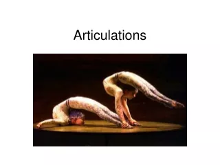
Articulations. junction of 2 bones MOTION OCCURS AT A JOINT -- NOT AT A LIMB i.e. elbow flexion NOT forearm flexion. Classification of joints. Synarthroses - fibrous joint with little or no movement Amphiarthroses - cartilaginous joints with some motion

E N D
Articulations • junction of 2 bones • MOTION OCCURS AT A JOINT -- NOT AT A LIMB • i.e. elbow flexion NOT forearm flexion
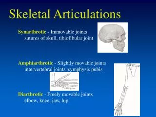
Classification of joints • Synarthroses - fibrous joint with little or no movement • Amphiarthroses - cartilaginous joints with some motion • Diarthroses - (aka synovial) - freely movable joint
Joint Classification • based on • number of axes of rotation • number of planes of motion • e.g. uniaxial -- 1 axis of rotation so 1 plane of motion
Ball and Socket = Triaxial e.g., flexion & extension internal & external rotation abduction & adduction Condyloid = Biaxial e.g., flexion & extension internal & external rotation
Pivot = uniaxial e.g., supination & pronation Hinge = uniaxial e.g., flexion and extension
Gliding = no axes ‘gliding between 2 flat bones’ Saddle = biaxial same as condyloid but greater ROM Ellipsoidal = biaxial e.g., flexion & extension abduction & adduction
Structure of Synovial Joint A - articular (hyaline) cartilage (1-7 mm) • smooth elastic tissue on ends of bone • 60-80% water • no blood supply • absorbs shock, distributes force and provides a low friction surface
Structure of Synovial Joint B - fibrous capsule • very fibrous collagen tissue used to hold bones together C - synovial membrane • lines the joint cavity • secretes synovial fluid to lubricate and provide nutrition NOTE: B & C combine to form the articular capsule or joint capsule
Structure of Synovial Joint D - ligaments • connect bone-to-bone • usually restrict ROM at a joint • tendons (not shown) • connect muscle-to-bone A* - Joint cavity
Other Structures of Synovial Joints Olecranon bursa • bursa • small capsules lined with synovial membranes • reduces friction between other structures in the joint • tendon sheaths • fascia surrounding tendon to reduce friction between tendon and surrounding structures Digital synovial sheath
Other Structures of Synovial Joints articular fibrocartilage • different from articular cartilage • takes the form of a fibrocartilaginous disc or partial disc • distributes load over joint surface • improve fit of articulating surfaces • limit slipping of one bone relative to other • protect periphery of articulation • lubricate articulation • absorb shock
close-packed vs. loose packed close packed position • maximum contact area • minimum mobility • maximum stability
Bony Stability (cont.) • amount of contact area
Joint Stability - Connective Tissue • ligamentous support
Properties of Connective Tissue • elasticity • ability to return to normal state after stretch • elastic limit • stretch beyond this limit will cause permanent damage • plasticity • stretched too far such that does not return to its normal state • ligament sprain (worse than bone fracture)
elastic limit Sprains occur in this region plastic load elastic deformation (length) Exercise will help increase the loads a ligament or tendon can sustain Sprains result in decrease of joint stability
Joint Stability - Muscles • muscular arrangement • ability of muscle to provide support • muscle fatigue • cruciate rupture more likely when muscle is fatigued
Mobility • degree to which an articulation is allowed to move before being restricted by surrounding tissues • ROM a.k.a. flexibility
Stability v. Mobility • trade-off between stability and mobility • increase stability decrease mobility • vice-versa
Neuromuscular Response to Stretching • Sensory neurons provide feedback on the characteristics of the muscle or other tissues. 2 neuromuscular proprioceptors: MUSCLE SPINDLES & GOLGI TENDON ORGANS
Muscle Spindles • location: • interspersed throughout muscle belly • responds to: • muscle length • muscle velocity • causes: • autogenic facilitation • reciprocal inhibition
Stretch Reflex • The muscle spindle is responsible for the stretch reflex. • As a muscle is rapidly stretched, the muscle spindle responds by facilitation of the same muscle and inhibition of the antagonistic muscle. • This reflex can be seen in the patellar tendon tap.
Golgi Tendon Organ • location: • near the muscle-tendon junction • responds to: • muscle tension • causes: • autogenic inhibition • antagonistic facilitation Muscle Fibers GTO tendon
“My Little GTO” • possibly the critical determinant to maximal lifting levels in weight training • may also be responsible for uncoordinated responses in untrained individuals • response is adapted through training
BALLISTIC STATIC activate muscle spindles which elicits a stretch reflex if static position achieved slowly then can minimize muscle spindle response if held for sufficiently long period (~30s) then can elicit GTO response may result in tearing a muscle STATIC BETTER THAN BALLISTIC
ACTIVE STRETCH Spindle response: minimal if performed slowly GTO response: active stretch of hip extensors causes GTO to relax hip extensors and to activate the hip flexors motive force: actions of the hip flexors consequences: no negatives -- limited ROM limits possibility of injury and exercise antagonists
PASSIVE STRETCH Spindle response: minimal if performed slowly GTO response: passive stretch of hip extensors causes GTO to relax hip extensors motive force: external force consequences: no direct control of ROM thus may exceed physiological limits and induce muscle damage
Stretching • Proprioceptive Neuromuscular Facilitation • PNF • alternating contraction - relaxation of agonist & antagonist muscles • takes advantage of the response of the proprioceptors • e.g. hamstrings • passive static stretch of hams - relax • active maximal concentric action of hams - relax • repeat
Plyometric Training Plyometric training consists of exercises that rapidly stretch a muscle followed immediately by a contraction. They improve power output in the muscle by: Neurological Influences: rapidly stretching of the muscle, which excites the motoneurons via the stretch reflex. Structural Influences: involving elastic energy from the stretch-shortening cycle.
Arthritis • Refers to more than 100 different diseases that affect areas in or around joints. • The disease also can affect other parts of the body. • Arthritis causes pain, loss of movement and sometimes swelling. • Affects women more than men Source: Arthritis Foundation – www.arthritis.org
Osteoarthritis 20.7 million Mostly after age 45 Fibromyalgia 3.7 million Mostly women Rheumatoid 2.1 million Mostly women Juvenile Arthritis 285,000 Under age 17 Gout 2.1 million Mostly men Arthritis Spondylarthropathies 412,000 Juvenile Rheumatoid Arthritis (JRA) 50,000 Lupus 239,000 Source: Arthritis Foundation – www.arthritis.org
Osteoarthritis (OA), or degenerative joint disease, is one of the oldest and most common types of arthritis, characterized by the breakdown of the joint's cartilage. Cartilage is the part of the joint that cushions the ends of bones. Cartilage breakdown causes pain and joint swelling. With time, there will be limited joint movement. • Most commonly affects middle-aged and older people • Range from very mild to very severe • Affects hands and weight-bearing joints (e.g., knees, hips, feet and back). • OA is not an inevitable part of aging, although age is a risk factor • Obesity may lead to osteoarthritis of the knees • Joint injuries due to sports, work-related activity or accidents may be at increased risk of developing OA. Source: Arthritis Foundation – www.arthritis.org
Rheumatoid Arthritis (RA) – a systemic disease that affects the entire body. • Characterized by the inflammation of the membrane lining the joint, which causes pain, warmth, redness and swelling. • The inflamed joint lining, the synovium, can invade and damage bone and cartilage. • Inflammatory cells release enzymes that may digest bone and cartilage. • The involved joint can lose its shape and alignment, resulting in pain and loss of movement. • The disease usually begins in middle age, but can start at any age, and affects two to three times more women than men. Source: Arthritis Foundation – www.arthritis.org
Location of “Tender Points” Fibromyalgia syndrome is a condition with generalized muscular pain and fatigue that is believed to affect approximately 3.7 million people. • The name fibromyalgia means pain in the muscles and the fibrous connective tissues (the ligaments and tendons). The condition is known as a syndrome because it is a set of signs and symptoms that occur together. • Fibromyalgia mainly affects muscles and their attachments to bones. Although it may feel like a joint disease, it is not a true form of arthritis and does not cause deformities of the joints. Fibromyalgia is, instead, a form of soft tissue or muscular rheumatism. Source: Arthritis Foundation – www.arthritis.org
Use of Heat or Cold Helpful before and after exercise Many respond better to cold packs than to heat Rest More rest and less activity are needed during flares and the opposite is true during periods of improvement. Medicines (e.g., analgesics, NSAIDS, DMARDS, Disease Modifying Anti-Rheumatic Drugs) Exercise (see next slide) Surgery joint replacement Arthritis Treatments Joint Protection Careful use of joints to limit the pressure on the involved joint Simple and inexpensive devices available • Diet • Lack of vitamins associated with progression of OA of the knee • Connection between obesity and OA of the knee • Diet high in Omega 3 fatty acids may help reduce inflammation in RA • In general, people with arthritis are urged to maintain a balanced diet and stay close to their ideal weight. • Physical/Occupational Therapy • recommend and teach prescribed muscle strengthening and range-of-motion exercises • teach non-medication ways to control pain • suggest ways to make everyday and work activities easier Source: Arthritis Foundation – www.arthritis.org
Exercise • Proper exercises performed on a daily basis are an important part of arthritis treatment. • Exercise to help reduce weight can help prevent osteoarthritis in the knee. • Proper exercise helps build and preserve muscle strength, keep joints flexible and help protect joints from further damage. • Two categories of exercise: • Therapeutic -- Prescribed by a doctor, physical therapist or an occupational therapist. These exercises are based on individual needs and are designed to reach a certain goal. • Recreational -- Includes any forms of movement, amusement or relaxation that refreshes the body and mind. These exercises add to a therapeutic program, but do not replace it. • Three types of exercises: • Range-of-motion -- Moving a joint as far as it comfortably will go and then stretching it a little further. Range-of-motion exercises are designed to increase and maintain joint mobility that will decrease pain and improve function. • Strengthening -- Increases muscle strength to stabilize weak joints. These exercises use the muscle without moving the joint. • Endurance -- This type of exercise includes walking, swimming, bicycling, jogging, dancing and skiing. These dynamic forms of exercise increase endurance, whereas range-of-motion and strengthening do not. The most common risk in exercising is injury to joints and muscles. This usually happens from exercising too long or too hard, especially if a person has not been active for some time. Source: Arthritis Foundation – www.arthritis.org