Download
1 / 54
560 likes | 838 Vues
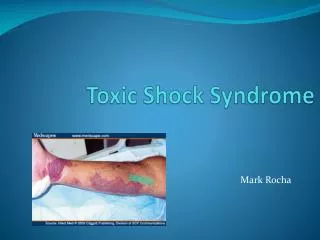
Shock Syndrome (Hypoperfusion Syndrome). Molnár Miklós Semmelweis University Institute of Pathophysiology 2 001. Shock- a rude unhinging of the machinery of life. Samuel Gross, 1872. First LeDran (1773) used this term (Fr. choc) to describe the

E N D
Shock Syndrome(Hypoperfusion Syndrome) Molnár Miklós Semmelweis University Institute of Pathophysiology 2001
Shock- a rude unhinging of the machinery of life. Samuel Gross, 1872 First LeDran (1773) used this term (Fr. choc) to describe the clinical characteristics of patients after severe gunshot trauma.
Definition • The term shock is used to describe complex pathophysiologic syndrome(s) arising from any multitude of causes. Shock usually results from a critical impairment of blood flow to vital organs and tissues and/or the ability of those tissues to utilize essential nutrients. The common denominator in all forms of shock is microcirculatory insufficiency
Common Cause of Shock 1. • Hypovolemic shock • Hemorrhage • Fluid loss • Gastrointestinal (e.g. vomiting, diarrhea) • Urinary (e.g. hyperglycemia, diabetes insipidus, diuretic therapy, postobstructive diuresis) • Skin (e.g. burn) • Internal sequestration (e.g. ascites)
Common Cause of Shock 2. • Cardiogenic shock • Myocardial failure • Left ventricular (e.g. ischemia, infarction, cardiomyopathy) • Right ventricular (e.g. infarction, pulmonary hypertension, cor pulmonale) • Arrhythmiák • Valvular regurgitation or stenosis • Ventricular septal rupture or free-wall rupture • Obstructive lesions • myxoma, pulmonary embolus, pricardial tamponade
Common Cause of Shock 3. • Distributive shock • Septic shock • Neurogenic shock (e.g. severe central nervous system depression, spinal cord injury) • Anaphylaxis • Adrenal cortical failure
Pathophysiology of Shock • Tissue hypoxia • Activation of protective mechanisms • Negative feed back mechanisms • Failure of the protective mechanisms • Positive feed back mechanisms • Over activation of the immune system • Multiple Organ Failure
Major Hemodynamic Determinants of Tissue Perfusion • Systemic arterial pressure Arterial Pressure=Cardiac Output x Total Vascular Resistance • Organ vascular resistance • Nutritional microcirculatory patency
Major Hemodynamic Determinants of Tissue Perfusion • Systemic arterial pressure • Total vascular resistance • Total arteriolar resistance, vascular muscle tone • tissue metabolites • neurohumoral factors • toxins • Blood viscosity
Cardiac output • Heart rate (brady- and tachyarrhythmias) • Stroke volume • preload (cardiac filling pressure and volume) • total circulating blood volume • distribution of blood volume • atrial contraction • diastolic filling time (heart rate) • Inotropic state • total functioning ventricular muscle mass • intrinsic (myocardial) control mechanism • extrinsic (noncardial) neurocirculatory control mechanisms • myocardial perfusion • myocardial oxygen demand • physiologic/pharmacologic depressant • humoral agents • Afterload • Aortic diastolic pressure • Ventricular size
Organ vascular resistance • Occlusive vascular disease • Local arteriolar and venular resistance • Neurogenic factors • Humoral factors • Local autoregulation • Nutritional microcirculatory patency • Precapillary sphincter tone • Postcapillary venular tone • Intracapillary aggregation of blood components • Capillary endothelial integrity
PRELOAD AFTERLOAD CONTRACTILE STATE Preload Contractile state Afterload Stroke volume Total circulating blood volume Distribution of blood volume body position intrapericardial pressure intrathorachal pressure venous tone Atrial Cotraction Aortic root diastolic pressure systemic vascular resistance arterial viscoelasticity aortic root blood volume Impedance Ventricular size Functioning ventricular muscle mass absolute mass oxygenisation Intrinsic/extrinsic neurohumoral mechanisms sympathetic nervous system sympathoadrenal axis circulating catecholamines parasympathetic nervous system Depressants hypoxia acidosis alkalosis drugs other circulating myocardial depressant factors
Pathophysiology of shock Stage I. Stage II. Stage III. Cellular membrane Injury Microcirculatory Failure Endothelial Damage Decreased perfusion Major End-Organ Dysfunction Low Cardiac Output or Vasodilatation Pathogenesis Compensated hypotension Decompensated hypotension Cellular Death ? Potentially reversibile shock Irreverzibile shock
Compensatory Mechanisms(negative feedback mechanisms) • Baroreceptor reflexes • Chemoreceptor reflexes • Cerebral ischemia • Reabsorption of tissue fluids • Endogenous vasoconstrictors • Renal conservation of water
Microcirculation and Transcapillary Exchange • Constriction of arteries and arterioles • Constriction of venues and venules • Capillary permeability and oncotic pressure edema
Exchange of Filtration-Reabsorption • Hemoconcentratio • Decrease of the Blood Volume • Stop of the Axial flow • rotation of red blood cells • blood sludging • Increased postcapillary resistance • Increased filtration
Effect of Long Lasting Hypoperfusion of Tissues • Accumulation of metabolites • lactic acid, vasodilators • Decreased Peripheral resistance • cerebral ischemia, cardiac ischemia - failure • Positive feedback mechanisms
Decompensatory mechanisms(positive feedback mechanisms) • Cardiac failure • Acidosis • Central nervous system depression • Alteration of blood clotting • Reticuloendothelial system
Cellular Membrane Integrity • Cellular Membran Injury • Hypoxia • Toxins, bacteriums, foreigner particles • Activation of Complement System • PLA2 • arachidonic acid • COX1/COX2 • LTB4, PGE2, TXA2, PGF1(primary mediators)
Mediators in Shock • Amins (histamine, serotonine) • Lipids • Eicosanoids, PAF • Proteolytic Cascade • Kinin-kallikrein • Complement system (C3a, C5a) • Clotting factors (XIII, vW) • Plasminogen - plasmin
Mediators in Shock 2. • Cytokines • Interleukins • IL-1, …. IL-15 … ILn • Tumor Necrosis Factor (TNF a,b) • Kolónia stimuláló faktorok (GCSF) • Growth Factors (FGF, TGF b) • Interferons (IF a, b, g) • Other polypeptides (fibronectin, chemokines) • Free Radicals • O2-, NO, lipid-, protein peroxides
Roles of Primary Mediators • Activation of endothelial cells • mediators (NO, ONOO-) • Activation of granulocytes • free radicals • lysosomal enzymes (myeloperoxidase, elastase) • migration • further activation • new mediator release (IL, TNF, PAF stb.) • inflammation
Decreased Cardiac Output Decreased arterial pressure Decreased systemic blood flow Decreased cardiac nutrition Decreased nutrition of tissues Intravascular clotting Decreased nutrition of vascular system Decreased nutrition Of brain Tissue ischemia Decreased vasomotor activity Increased capillary permeability Release of toxins Vascular dilatation Decreased Blood volume Venous pooling Cardiac depression Decreased venous return
Hypovolaemic Shock • Hemorrhage • Trauma, surgery, aneurysm rupture • Hemothorax, hematoma • Haemophylia, anticoagulants, thrombolytics • Exessive Fluid Loss from GI Tract • Vomiting, diarrhea -- especially infants and children • Urinary Tract Fluid Losses • Diabetes insipidus, diabetes mellitus, salt-wasting disorders, adrenocortical insufficiency, diuretics • Fluid Loss from the Skin • Excessive burn, skin inflammation (generalized exfoliative dermatitis) • Internal Sequestration of Fluid • Loss of volume into the interstitial space or body cavities. Chronic liver disease, acute pancreatitis, angioedema.
Cardiac Output Preload Effect of Hypovolaemia on Cardiac Output
I. 100 II. III. 80 IV. 60 V. 40 VI. 20 0 60 120 180 240 300 360 Course of Arterial Pressure in Dogs after Different Degrees of Acute Hemorrhage Arterial pressure (% of control) 0 Time(min)
Effect of Hemorrhage on Cardiac Output and Arterial Pressure 100 Arterial Pressure Cardiac Output and Arterial Pressure (% of normal) 50 Cardiac Output 0 0 20 40 50 % of Total Blood Removed
Baroreceptor Reflexes Cerebral Ischemia b1-receptors Increasing heart rate and cardiac output b2-receptors metabolism a-receptors vasoconstrictio (kidney, splanchnic area, muscle, adipose tissue) Renin-Angiotensin-System ADH/Vasopressin Compensatoris Mechanisms in Hypovolemic Shock
Consequences • Almost normal cerebral and cardial circulation (70-90 mmHg) • Circulation of other organs decrease -TPR • Skin - pale • Kidney - decreased diuresis • Muscle - weakness
Impaired Pump Function Myocardial Infarction Asthma cardiale Severe Acidosis Barbiturat intoxication Toxins of Septic Shock Valvular regurgitation or stenosis Septal Rupture Pericardial tamponade Pneumothorax Embolism Cardiogenic Shock
Cardigenic shock Cardiac Output Preload
Hyperdinemic shock • Septiko-toxic shock • Normovolemia • Normotension • Decreased TPR (generalized vasodilatation) • Increased Cardiac Output (2-3 fold) • Redness, fever • Increased Heart rate
Hyperdinamic shock • Accumulation of lactic acid • Changes in amino acid metabolism • tyrosine - octopamine inhibits the a-receptors • Increased production of NO • cytokines, endotoxin - iNOS • Relative oxygen deficits -- cardiac failure
Distributive shock Cardiac Output Preload
Reperfusion • Hypoxia • large artery (a femoralis, a mesenterica sup.) • Reperfusion • liberation of mediators
Reperfusion Injury • Severe hypoxia/anoxia • Tissue injury (endothel cells etc.) • Early reperfusion: No reflow (capillaries); Reflow paradox (postcapillar venules) • Late reperfusion • Inflammation
Delayed Consequences of Reperfusion • Inflammatoric reactions • Local • Diffuse • Compensatoric mechanisms • Apoptosis • Tendency for regeneration
Delayed Consequences of Reperfusion • Hemorrhagic Necrosis • Hemorrhage • Fluid loss
Causes of Tissue demage • Ca2+influx • Enzyme activation (PLA2, XOR, iNOS stb.) • Free radicals (XO, XD) • Primary mediators • Histamine, eicozanoids, NO, peptides, Etc. • Cell-cell interaction • Endothel layer –PMN mediator release
Septico-toxic shock • Hyperemic Stage • Inpaired utilization of oxygen • Bacterial sepsis • Endotoxin, severe burn, necrotic tissue demage
Anaphyxis • Sensitized individual exposed to antigen triggers an IgE-mediated activation of mast cells. • Decreased sympatetic inervation of vasculature • Drugs (penicillin), food (eggs, white nuts, seafood) • Direct smooth muscle relaxation • endotoxin • endotoxin induced cytokine release
Trauma multiple Direct Tissue Demage Inflammation Hemorrhage Hypovolemic shock hypoxia Tissue injury Inflammation Infection Inflammation Sepsis
A máj szerepe a sokkban 1. • A hepatocytákra hatnak: NO, immunsejtek, hormonok stb. • Fokozott aminósav kínálat (alanin, glutamin) • Fokozott tejsav-, piroszőlősav kínálat • Ammónia-nitrogén -- karbamid szint emelkedik • Csökken a fenilalanin és a tirozin felvétele, a leuciné nem változik (leu/tyr, leu/phe)
A máj szerepe a sokkban 2. • Hyperglykaemia • glukoneogenezis • tejsav, piroszőlősav, alanin Kezdetben fontos tápanyag, később azonban a túlműködést és a baktériumok szaporodását segítik elő
Akut fázis fehérjék fokozott aminosav kínálat IL-1, IL-6, TNF Kuppfer sejtek (RES) egyéb C-reaktív protein gyulladást serkentése granulocyta migráció fokozása phagocyták kitapadása komplement aktiváció a1-antitripszin antiproteolítikus Cöruloplazmin, a2 -makroglobulin, transzferin szabadgyökfogók, antiproteázok A máj szerepe a sokkban 3.
A máj szerepe a sokkban 4. • Alvadási rendszer • Komplement rendszer • Fibrinolítikus rendszer A fibrinogén szint traumás és szeptikus sokkban a folyamat súlyosságát jelzi.
Az izomszövet reakciója sokkban • Hemodinamikai szerep (A testtömeg 30-40%) • Anyagcsere termékek • A filtráció nő -- vértérfogat csökken • Citokinek (TNF, IL-1, IL-6 és GIF) • A TNF gátolja a piruvát dehidrogenázt • glukóz felvétele nő, csökken a glukóz oxidációja • piroszőlősav-, tejsav leadás nő • fehérjék fokozott lebontása (leucin) -- ketonsavak • piroszőlősav + aminócsoport: alanin A plazma alanin koncentrációja nő
Az endokrin rendszer szerepe sokkban • Adrenalin -- vércukorszit emelkedés • Kortizol -- fokozott fehérjebontás • Inzulin és a glukagon mennyisége nő • (a keringésbe kerülő citokinek miatt)
A lipidanyagcsere változásai sokkban • Fokozott tejsav, piruvát, alanin, ketonsavak TCA ciklus acetil-KoA • Inzulin -- malonil-KoA -- zsírsavszintézis -- lipidek felépülése • Izomban: zsírsavégetés, glukóz oxidációja helyett
A tüdő kóros elváltozása sokkban • Immunválaszok túlaktiválódása • Adult Respiratory Distress Syndroma (ARDS) a MOF részeként • 70%-os mortalitás • alveoláris folyadékgyülem • tromboxán, endotoxinok, az aktiválódott immunsejtek növelik a kapilláris membrán permeábilitását • fehérje lép ki -- ozmotikus gradiens csökken • alveolus membrán epitel sejtjei károsodnak • romlik a gázcsere (diffúziós út nő, légzőfelület csökken, AV söntök megnyílnak) • arteriás pO2 csökken -- Légzési elégtelenség