Download
1 / 25
250 likes | 440 Vues
The third annual International Neurosurgery Conference. Space‐occupying Cerebellar Infarcts: A Review. Luis Rafael Moscote-Salazar. MD Kalil Kafury-Bennedeti. MD Rubén Sabogal-Barrios. MD. UNIVERSIDAD DE CARTAGENA Cartagena de Indias, COLOMBIA 2007. EPIDEMIOLOGY.

E N D
The third annual International Neurosurgery Conference Space‐occupying Cerebellar Infarcts: A Review Luis Rafael Moscote-Salazar. MD Kalil Kafury-Bennedeti. MD Rubén Sabogal-Barrios. MD UNIVERSIDAD DE CARTAGENA Cartagena de Indias, COLOMBIA 2007
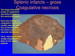
EPIDEMIOLOGY Cerebellar infarcts are not uncommon: they account for 2-4% of all strokes . Proportions 4-5 times higher than for cerebellar haemorrhages.
CLINICAL FEATURES AND PATHOPHYSIOLOGICAL MECHANISMS • Thecerebellumissuppliedbythreemainarteries, each of wichalso has a correspondingterritory in thebrainstem. • Cerebellarinfarctsinvolvingthe posterior inferior artery (PICA) and the superior cerebellarartery (SCA) are mostcommon, whereasinfarctsinvolvingthe anterior inferior cerebellarartery (AICA) are rare.
Posterior inferior cerebellarartery (PICA) infarcts • The PICA arisefrom vertebral artery, and divides into medial (mPICA) and lateral (lPICA) branch. ThemPICAsometimespartlysuppliesthe lateral medullaoblongata, butmostoftenthisregionissuppliedbybranchesoriginatingdirectlyfrom vertebral artery. • Infarct in themPICA are characterizedbyvertigo, dizziness, truncal ataxia, axial lateropulsion and nystagmus.
PICA infarcts are most often caused by large artery occlusive disease in the vertebral arteries, whereas cardiac embolism account for a 20% of infarcts.
Anterior Inferior cerebellarartery (AICA) Infarcts • AICA infarcts are almostalwaysaccompainiedbybrainstemsignsfromlowerpons. AICA infarctshavebeenconsideredveryrare, buttheirfrequencymighthavebeenunderstimatedbecausesomehaveprobablymisdiagnosed as lateral medullaryinfarcts. • AICA infarcts are usuallyduetolargearterydisease in thelower basilar artery.
Superior cerebellarartery (SCA) Infarcts • The SCA suppliesthelaterotegmentalportion of the rostral ponsincludingthe superior cerebellarpeduncle, spinothalamictract, lateral lemniscus, descendingsympathetictract and root of thecontralateralIVthcranialnerve. • The SCA has twobranches: the medial branch (mSCA) and the lateral branch (lSCA) supplyingthedorsomedial and anterolateralareas, respectly.
SurgicalTreatment Rapidly progressive cerebellar swelling with acute hydrocephalus, brain stem compression, and death is a feared complication of cerebellar infarct. Careful monitoring of patients with cerebellar infarcts, in particular those with large PICA infarcts and in multiple posterior circulation infarcts, for 3-4 days is therefore essential.
The surgical management of space occupying cerebellar infarcts has been much debated, partly reflecting the lack of randomised clinical trials.
Surgical Management Suboccipital craniectomy and removal of necrotic tissue, envolving hydrocephalus (for which external ventricular drainage may be attempted ) or concomitant irreversible brain stem infarction (for which no surgcial procedure is likely to be helpful).
OUTCOME OF SURGERY The outcome of surgery depends much on wheter there is an brainstem infarct. There is no evidence for the use of thrombolytic therapy in isolated cerebellar infacrt.
Patient W.M. • History of Present Illness: • 34 year old male • Long history of headaches • Presented with 8 days of: • Bitemporalheadache progressing to • Bifrontal headache • Somnolence • Altered mental status • Nausea/vomiting • dizziness • No fevers, chills • No history of trauma
Patient W.M. • Past Medical History: • Otherwise unremarkable past medical history • Medications: • None • Allergies: • None Known • Social History: • No tobacco, drug, or alcohol use
Patient W.M. • Physical Exam • Mental Status • Patient somnolent, • Oriented inconsistently to name only • Cranial Nerve Exam • Extraocular movements intact • Cranial Nerves otherwise intact
Patient W.M. • Motor exam • Anormaltone • Follows simple commands intermittently • Diffusely weak in all extremities • Sensory Exam • Sensation intact to light touch in all extremities • Reflexes • Reflexes 2+, symmetrical • No Hoffman’s sign • Toes downgoing • Cerebellar/Gait exam • Mild dysmetria bilaterally on finger-nose test • Gait Deferred
CONCLUSIONS In patients with deteriorating cerebellar infarcts a repeat neuroimaging Study usually identifies the cause of worsening and is very helpful usually Identifies the cause of worsening and is very in guiding the use of Surgical intervertions. Space‐occupying Cerebellar Infarcts is a Neurosurgical Pathology Close monitoring for 3-4 days is warranted in cases of large cerebellar infarcts and multifocal posterior circulation ischaemia. Neuroimaging with MRI/dw-MRI/MR-angio should be liberally used in suspected cerebellar infarcts, because findings usually influence therapy.