Download
1 / 33
350 likes | 959 Vues
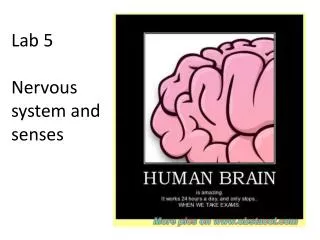
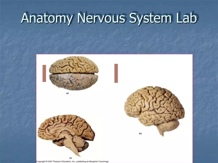
Anatomy Nervous System Lab. Divisions of the Nervous System. Central nervous system – brain & spinal cord Peripheral nervous system – cranial nerves & spinal nerves. The Brain. Brain stem medulla oblongata (M.O.) pons midbrain Diencephalon thalamus hypothalamus

E N D
Divisions of the Nervous System • Central nervous system – brain & spinal cord • Peripheral nervous system – cranial nerves & spinal nerves
The Brain • Brain stem • medulla oblongata (M.O.) • pons • midbrain • Diencephalon • thalamus • hypothalamus • mamillary bodies • epithalamus (pineal gland) • Cerebrum • Cerebellum Cerebrum T P P H midbrain M Cerebellum pons m.o.
The Brainstem • medulla oblongata (M.O.) • pons • midbrain – • cerebral peduncle • cerebral aqueduct of midbrain • corpora quadrigemina • superior colliculi • inferior colliculi pons Cerebral peduncle m.o. Cerebral aqueduct of midbrain
The Diencephalon • thalamus • hypothalamus • mamillary bodies • connects to pituitary gland via infundibulum • epithalamus (pineal gland) T P H M infundibulum
The Diencephalon Intermediate mass of Thalamus Pineal gland Hypothalamus Infundibulum Pituitary gland
Cerebrum convolutions gyrus sulcus (separated from cerebrum by transverse fissure) 4 lobes per hemisphere- frontal, parietal, temporal, occipital
corpus callosum septum pellucidum fornix The Cerebrum
gray matter “folia” • white matter “arbor vitae” • separated from cerebrum by transverse fissure • 2 hemispheres connected by “vermis” The Cerebellum Transverse fissure
The Spinal Cord • Begins at foramen magnum & ends at L2 vertebral level by forming conus medularis • Has 2 thickened areas- cervical enlargement - supplies nerves to upper extremity • lumbar enlargement - supplies nerves to lower extremity • Made up of 31 spinal cord segments
Posterior median sulcus Posterior column Posterior gray horn - sensory Central canal Lateral column Lateral gray horn (T1-L2, S2-S4) - autonomic Anterior gray horn - motor Anterior column Anterior median fissure Cross Sectional Anatomy of the Spinal cord segments Dorsal root Dorsal root ganglion Gray commissure Spinal nerve Ventral root
The Spinal Cord & Spinal Nerves • Each spinal cord segment gives off a pair of spinal nerves • each spinal nerve forms from union of dorsal/ventral root of spinal cord segment & exits between vertebra at IVF • 8 pair cervical spinal nerves – 1st cervical nerve exits between occipital bone & C1, 8th cervical nerve exits the IVF between C7-T1 • 12 pair thoracic spinal nerves • 5 pair lumbar nerves • 5 pair sacral nerves • 1 pair coccygeal nerves
Below the conus medularis, spinal nerves must angle downward (in the subarachnoid space) before exiting their IVF. These spinal nerves make up the cauda equina Cauda equina
The Central Nervous System CNS well protected by bones (cranial & vertebrae), CT meninges, & cerebrospinal fluid (CSF) Meninges – Connective tissues that surround and protect the brain and spinal cord (CNS) • 3 layers: • Dura Mater • Arachnoid • Pia Mater
Meninges Dura Mater – tough, fibrous outer layer • 2 layers thick around brain with creation of dural sinuses between layers; dural folds into fissures of brain
Superior sagittal sinus Falx cerebri Inferior sagittal sinus Tentorium cerebelli Straight sinus Confluence of sinuses Transverse sinus Sigmoid sinus Dural folds and dural sinuses
Dural folds and dural sinuses Superior sagittal sinus Falx cerebri Inferior sagittal sinus Straight sinus Confluence of sinuses Transverse sinus
Meninges- Dura mater • 1 layer around spinal cord with epidural space external
Meninges Arachnoid– “spidery” web-like middle layer
Pia Mater – delicate, thin inner layer; • filum terminale - extension of pia mater extends from tip of cord to coccyx to anchor cord in place; • denticulate ligaments anchor cord laterally
Meninges Subarachnoid space – between arachnoid & pia mater; contains cerebral spinal fluid (CSF)
Cerebrospinal Fluid (CSF) • clear, colorless fluid formed by filtration of blood plasma by choroid plexuses within ventricles of the brain. • functions in protection of CNS, support, nutrient supply, waste removal
CSF Circulation Lateral ventricles (in cerebral hemispheres) interventricular foramen third ventricle (in diencephalon around thalamus) cerebral aqueduct of midbrain fourth ventricle (between pons/cerebellum) subarachnoid space & central canal of SC Lateral ventricle (behind septum pellucidum) Third ventricle Aqueduct of midbrain Fourth ventricle
Reabsorption of CSF through arachnoid granulations (arachnoid villi) of dural sinuses (superior sagittal sinus) into cerebral veins
Cranial Nerves • 12 pairs of nerves that connect to the brain; provide motor, sensory &/or autonomic (parasympathetic) function
Spinal nerves & Plexuses (Intercostal nerves) (Common peroneal nerve)