Download

1 / 17
180 likes | 315 Vues
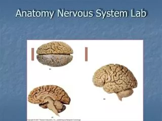
Lab 5 Nervous system and senses. Nervous System Two primary divisions of the nervous system: Central nervous system ( CNS) - consists of the brain and spinal cord Peripheral nervous system ( PNS) - consists of all neural tissue outside of the CNS .

E N D
Lab 5 Nervous system and senses
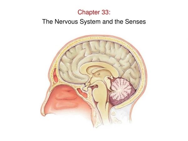
Nervous System Two primary divisions of the nervous system: Central nervous system (CNS)- consists of the brain and spinal cord Peripheral nervous system (PNS)- consists of all neural tissue outside of the CNS
The CNS contain both gray matter and white matter gray matter - areas of CNS dominated by nerve cell bodies and unmyelinated axons white matter - areas of CNS dominated by myelinated axons
The CNS is are covered by meninges (membranes) for protection and cushioning dura mater - tough outer layer arachnoid mater - spider-like middle layer pia mater - transparent inner layer
CNS also contains cerebrospinal fluid - fluid bathing the surfaces of CNS • fills ventricles and subarachnoid space • secreted by choroid plexus (complex interwoven network of peripheral nerves) • functions: -protection, drains unwanted substances away from brain – helps maintain homeostasis
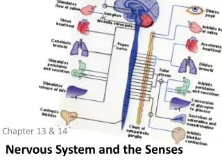
Autonomic Nervous System – monitors and adjusts body systems to maintain homeostasis – involuntary (think automatic) Sympathetic – stimulates metabolism, increased heartbeat, and elevates mental alertness to allow the body to cope with a stressful situation. Prepares body for “fight” or “flight” action. Widely distributed throughout the body. Parasympathetic – conserves bodyby promoting sedentary activities, such as “rest and digest” functions. Limited to the head, neck, and selected viscera.
Relationships Between The Sympathetic And Parasympathetic Divisions Dual Innervation – most organs are innervated by both divisions thru plexuses with opposing effects
Special Senses: Ear • Functions of ear: hearing and balance • static equilibrium (linear acceleration) • dynamic equilibrium (rotational acceleration)
Regions of the ear: outer ear - pinna, external auditory canal/meatus, tympanic membrane (ear drum), captures sound
middle ear – ossicles: 3 bones - malleus/hammer, incus/anvil, stapes/stirrup, auditory/eustacian tube, amplifies sound
inner ear - network of canals (membranous labyrinth) with receptors, balance and sound detection
Regions of the eye: fibrous tunic - outermost region, epithelial tissue (integument) (NOT vascularized) sclera - opaque white, function is support cornea - transparent, function is protection
vascular tunic - middle region (vascularized) iris - pigmented, contains smooth muscle that contract to change the diameter of the central opening of the iris pupil - regulates the amount of light entering the eye. choroid - dark in color:many blood vessels, supplies nutrients + oxygen to retina ciliarybody - attached to lens and can change shape of lens to focus
neural tunic orretina - neural inner region with two primary layers • outerpigmented layer - absorbs light • innerneural layer – contains a photoreceptors layer • rods- b/w or dim light • cones– color or bright light • bipolar layer - supporting cells • ganglioniclayer - neurons that perform preliminary processing and integration of visual information
macular zone - "yellow spot" - no rods in this section of photoreceptive layer, the highest concentration of cones is found in the center of the macular zone is the fovea optic disc (blind spot) - axons from the ganglionic layer converge here and penetrate the wall of the eye and proceed toward the brain via the optic nerve.