Download
1 / 33
330 likes | 389 Vues
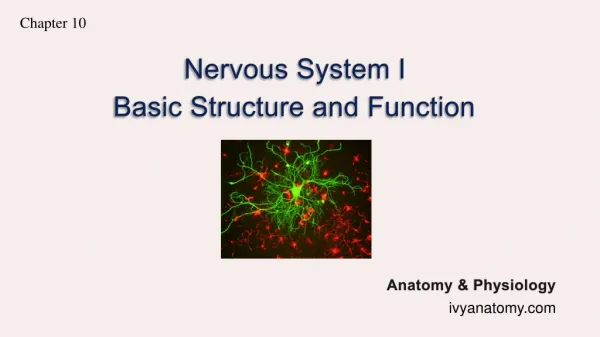
Nervous System I Lab 5 ( rev 3-11). The CNS consists of the brain and spinal cord is the integrating and command center of the nervous system it interprets incoming sensory information and produces motor responses.

E N D
Nervous System I Lab 5 ( rev 3-11) • The CNS consists of the brain and spinal cord • is the integrating and command center of the nervous system • it interprets incoming sensory information and produces motor responses BIO lab 102 Nervous I- Lab 5
The peripheral nervous system is the part of the nervous system outside the brain and spinal cord (the CNS) • it contains the communication lines that link all parts of the body to the CNS • consists mainly of nerves that extend from the brain and spinal cord • the Peripheral NS has 2 functional subdivisions • the sensory or afferent division carries impulses TO the CNS and keeps the CNS informed of events going on inside and outside of the body BIO lab 102 Nervous I- Lab 5
Nervous System I Lab 5 • The motor or efferent division which carries impulses FROM the CNS • This division enables us to respond to stimuli • The Motor Division can be further subdivided into: • the Somatic or Voluntary nervous system • carries impulses from the CNS to skeletal muscles thus allowing us to consciously control our skeletal muscles BIO lab 102 Nervous I- Lab 5
The Autonomic nervous system (ANS), is also referred to as the involuntary nervous system • this division regulates the activity of smooth muscles, cardiac muscles, and glands • Is further sub-divided into the • the SYMPATHETIC NERVOUS SYSTEMwhich mobilizes body systems during emergency situations • the PARASYMPATHETIC NERVOUS SYSTEMwhich primarily conserves our energy BIO lab 102 Nervous I- Lab 5
Composition of the Nervous System • The Nervous System is composed of 2 types of tissue • Neurons are the conductive cells which transmit messages • Neuroglia are the supporting cells of the nervous system BIO lab 102 Nervous I- Lab 5
Neurons • Are the specialized cells which conduct nerve impulses • All neurons have • dendrites (receive stimuli) • a cell body (contains the nucleus) • an axon (carries impulses) • Some neurons have a myelin sheath around the axon BIO lab 102 Nervous I- Lab 5
the myelin sheath • is a fatty wrapping around the axon which provides insulation to the axon • it speeds impulse transmission • is formed from Schwann Cells in the peripheral nervous system and Oligodendrocytes in the CNS • Between neighboring Schwann cells are short, uninsulated gaps called Nodes of Ranvier BIO lab 102 Nervous I- Lab 5
Neurons can be classified based on structure or function:Structural Classification Unipolar Neurons • have a single process which emerges from the cell body • this process divides into a proximal and distal branch • One branch behaves as an afferent branch and the other behaves as an efferent branch • All unipolar neurons are sensory BIO lab 102 Nervous I- Lab 5
Bipolar Neurons • have 2 processes emerging from a round cell body • processes extend from opposite sides of the cell body • found only in some of the special sense organs where they act as receptor cells (i.e. located in the retina [eye], in the ear and olfactory mucosa) Multipolar Neurons • have 3 or more processes • are the most common neuron type in humans and major neuron type in the CNS BIO lab 102 Nervous I- Lab 5
Classification Systems: Structural Classification Interneurons or Association Neurons • are a multipolar neuron • located in the CNS • conduct impulses within the CNS • are the connecting link between sensory and motor neurons BIO lab 102 Nervous I- Lab 5
Functional Classification • Sensory (Afferent) Neurons • carry (sensory) information from receptors to the CNS • Most are unipolar; a small number are bipolar • Motor (Efferent) Neurons • carry messages away from the CNS to the muscles and the glands • are multipolar BIO lab 102 Nervous I- Lab 5
Nervous System I Lab 5 Neuroglial or Glial Cells • are non-nervous tissue cells which provide a supportive scaffolding, protect and nourish neurons • Astrocytes anchor the neurons to nearby capillaries • Ependymalcellsare ciliated cells that move cerebrospinal fluid around the brain and spinal cord • Microglia are phagocytic cells for the nervous system. They destroy bacteria and damaged tissues near the neurons. • Oligodendrocytes form the myelin sheath in the CNS • Schwann cells form the myelin sheath in the PNS BIO lab 102 Nervous I- Lab 5
Impulse Transmission This is primarily a lecture topic; we will quickly go over this. • An impulse arrives at the dendrite • When the impulse is strong enough, it depolarizes the membrane and the impulse is transmitted along the axon • When the impulse reaches the axon terminals, the information needs to be converted to another form of energy in order for the information to be transmitted to its target (i.e. a muscle, a gland, or another neuron) • A chemical, called a neurotransmitter, is released which allows the impulse to jump the synapse, or space, between the 2 cells BIO lab 102 Nervous I- Lab 5
The Brain and the Spinal Cord The CNS controls and processes all information received by the body. • Protection of the CNS • Bone protects it from physical injury • the brain is covered by the skull • the spinal cord is surrounded by the vertebrae BIO lab 102 Nervous I- Lab 5
Meninges--3 protective membranes of connective tissue enclose the brain and spinal cord • Dura mater is the outermost and strongest layer • Arachnoid mater is the middle layer; it has spidery extensions which secure it to the innermost layer. • Pia mater is the innermost layer and clings tightly to the CNS. BIO lab 102 Nervous I- Lab 5
Cerebrospinal fluid (CSF): is found within and around the brain and spinal cord. It • functions as a liquid shock absorber • serves as a blood-brain barrier by isolating the CNS from infections • aids in providing nutrients for cells and removing waste products from cells • by floating the brain, the CSF effectively reduces brain weight by 97% and prevents the brain from crushing under its own weight BIO lab 102 Nervous I- Lab 5
CSFNervous System I Lab 5 • CSF is formed from blood plasma • it is made in the choroid plexuses in the ventricles of the brain • much of the fluid is found in the subarachnoid space BIO lab 102 Nervous I- Lab 5
Gross Anatomy of a Nerve • Nerves consist of parallel bundles of axons wrapped by various layers of connective tissue. • From the inside to the outside, • each axon is covered by endoneurium, a delicate layer of connective tissue. It also covers the myelin sheath and neurilemma. • groups of axons are bound into bundles called fascicles. • the coarse layer of connective tissue which wraps the fascicles is the perineurium. • the epineurium surrounds the fascicles. It is a tough, fibrous covering. BIO lab 102 Nervous I- Lab 5
Spinal Cord • The spinal cord is located in the vertebral column. • It is approximately the width of the thumb except at the cervical and lumbar areas. • Cervical enlargement is where the nerves for the shoulder and arms enter and exit the cord. • Lumbar enlargement is where the nerves for the legs enter and exit the cord. BIO lab 102 Nervous I- Lab 5
Spinal CordNervous System I Lab 5 • The spinal cord does not extend to the end of the vertebral column. It ends at the level of L2(the area between the first and second lumbar vertebrae). • The cord ends in a tapering cone shape which is called the conus medularis. • The lumbar and sacral nerves angle sharply downward and travel through the vertebral canal before they exit through intervertebral foramina. • The collection of these nerves is called the cauda equina because they resemble a horse’s tail. BIO lab 102 Nervous I- Lab 5
The outer portion of the spinal cord consists primarily of bundles of axons called nerve tracts. • These axons are usually myelinated and so they have a white appearance and are called “white matter.” • White matter is made up of ascending (sensory) and descending (motor) nerve tracts of myelinated axons • Near the center of the cord is an area called the gray matter. It is occupied primarily by cell bodies, dendrites and axons of CNS neurons (which are not myelinated). • Is in the shape of an “H”; this is where all synapses between sensory, association, and motor neurons takes place. BIO lab 102 Nervous I- Lab 5
Spinal Cord Anatomy-Other Things to Know • The Central Canal is where the CSF circulates. • The Ventral (or anterior) Root is where all MOTOR axons exit from the spinal cord. • The Dorsal (or posterior) Root is where all SENSORY axons enter the spinal cord. • The Dorsal Root Ganglion is where all sensory nerve cell bodies are located. BIO lab 102 Nervous I- Lab 5
Spinal NervesNervous System I Lab 5 • Dorsal and ventral roots join to form Spinal nerves. These are called mixed nerves because they have axons of both sensory and motor nerves. • Spinal nerves unite with other nerves to form a plexus. • There are 8 pairs of cervical nerves (C1-C8). • Most go to the cervical plexus • except C8 which goes to the brachial plexus. • Major nerve emerging from the cervical plexus is the phrenic nerve (innervates the diaphragm). BIO lab 102 Nervous I- Lab 5
Spinal NervesNervous System I Lab 5 • The brachial plexus is formed from C8, T1and T2. • Major nerves to emerge are the radial and ulnar nerves • these innervate many of the muscles of the forearm and hand • The 12 pairs of thoracic nerves (T3 through T12) do not go to a plexus. • They innervate the intercostal muscles used for breathing. BIO lab 102 Nervous I- Lab 5
Spinal NervesNervous System I Lab 5 • There are 5 pairs of lumbar nerves, L1 through L5. Most go to the lumbar plexus. • Major nerve is the femoral nerve which innervates the anterior thigh muscles and the skin covering them. BIO lab 102 Nervous I- Lab 5
Spinal NervesNervous System I Lab 5 • There are 5 pairs of sacral nerves, S1 through S5. These go to the sacral plexus. • Major nerve is the sciatic nerve which innervates the posterior thigh muscles and its skin covering. • The sciatic nerve is the largest nerve in the body. • The coccygeal nerve does not go to a plexus. • It innervates genitoanal structures. BIO lab 102 Nervous I- Lab 5
The Reflex Arc Many of the body’s control systems belong to a general category known as reflexes. • A reflex is a rapid, predictable motor response to a stimulus. • It is automatic, involuntary, and protective. • Both the spinal cord and the brain are reflex centers. BIO lab 102 Nervous I- Lab 5
Reflex ArcsNervous System I Lab 5 Reflex arcs include the following components. 1. The receptoris the where the stimulus begins. 2. The sensory neuron transmits the afferent impulses to the CNS. 3. An association neuron receives the information and causes an instantaneous impulse to be transmitted to a motor neuron. BIO lab 102 Nervous I- Lab 5
Reflex ArcNervous System I Lab 5 4. The motor neuron sends an impulse to the effector organ (organ which will cause a response). 5. The effectoris the muscle or gland that responds in a characteristic way. This allows us to respond to a stimulus instantly and without thinking. BIO lab 102 Nervous I- Lab 5
More Reflex ArcsNervous System I Lab 5 • The Babinski Reflex is an extremely important reflex. • It is normal in newborns to 6-12 months; it is abnormal in older children and adults. • Presence of this reflex indicates damage to the motor cortex of the CNS. • To test for this reflex, stroke the lateral aspect of the foot starting from the heel, up to and across the ball of the foot and end at the big toe. • An abnormal response is dorsiflexion of the big toe with extension and fanning of the other toes. BIO lab 102 Nervous I- Lab 5
REMINDER, page 1 of 2: 1. There are slides on the desks relating to nerve cells. We will not be using them today since you have already seen examples of this in the earlier labs when we were looking at cells. Focus on the models around the room. 2. Learn the 3 types of neurons and structure of the neuron on models and diagrams (no slides). BIO lab 102 Nervous I- Lab 5
REMINDER, page 2 of 2: 3. Learn the structure of the spinal cord, plexuses and spinal nerves on models and diagrams. 4. Learn the parts of the reflex arc. 5. Do the patellar reflex (skip ankle and Babinski reflexes). Do NOT hit the patella--feel for the space under it and tap in this space. Get permission from your partner prior to using the reflex hammer. BIO lab 102 Nervous I- Lab 5