Download
1 / 226
2.42k likes | 3.11k Vues
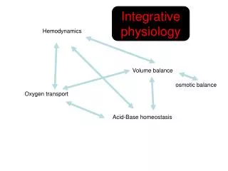
ECHOCARDIOGRAPHY AND HEMODYNAMICS REVIEW ECHO II. Susan A. Raaymakers , MPAS, PA-C, RDCS (AE,PE) Coordinator of Radiologic and Imaging Sciences - Echocardiography Grand Valley State University, Grand Rapids, Michigan raaymasu@gvsu.edu. Anatomy. Right Atrium SVC, CS, IVC Smooth walled

E N D
ECHOCARDIOGRAPHY AND HEMODYNAMICS REVIEWECHO II Susan A. Raaymakers, MPAS, PA-C, RDCS (AE,PE) Coordinator of Radiologic and Imaging Sciences - Echocardiography Grand Valley State University, Grand Rapids, Michigan raaymasu@gvsu.edu
Anatomy • Right Atrium • SVC, CS, IVC • Smooth walled • Derived from embryonic right atrium called the sinus venosus • Right atrial appendage • Sulcus terminalis • Posterior external ridge that extends vertically from the SVC to the inferior vena cava • Pectinate muscles
Name this complication of coronary artery disease • acute inferoseptal and inferior myocardial infarction resulting in ventricular septal defect
1. ______ 2. ______ 3. ______
Name the Wall Segments AND Typical Perfusing Coronary Artery Apical Cap LAD
Name the Wall Segments AND Typical Perfusing Coronary Artery Basal Anteroseptum LAD
Name the Wall Segments AND Typical Perfusing Coronary Artery Apical Cap LAD
Name the Wall Segments AND Typical Perfusing Coronary Artery Mid Anteroseptum LAD
Name the Wall Segments AND Typical Perfusing Coronary Artery Basal Anteroseptum LAD
1. _________ 2. _________ 3. ________
What is this complication of myocardial infarction called? When does it occur? • Dressler’s Syndrome: delayed form of pericarditis: an immunologic reaction • Occurs one to 12 weeks post MI • Symptoms: fever, pleuropericaridial pain, malaise • Cardiac tamponade is rare
What is polyarteritis? (Also called Kussmaul's disease, periarteritis nodosa) • Systemic inflammation and necrosis occurring in medium-sized or small arteries. • Kidneys, heart, liver, GI tract, pancreas, testes, skeletal muscular system, central nervous system (CNS), and skin are involved.
1. _________ 2. _________ 3. _________
Ischemia results in narrow of __________ percentage of luminal cross sectional area. This causes blood flow to become inadequate to meet demand with exercise, mental stress or pharmacologic interventions. >70%
T/F This spectral Doppler image of mitral regurgitation is consistent with a reduced dP/dt and is consistent with increased left ventricular end-diastolic pressure. True. The MR is quickly leaving the LV causing the LA to quickly increase in pressure.
Name four risk factors for coronary artery disease. • Increased LDL • Smoking • Diabetes • Hypertension • Genetics (hereditary) • Type “A” personalities • Aging • Obesity • Sedentary lifestyle • Chronic stress
Name this complication of coronary artery disease • Portion of papillary muscle seen in transesophageal echocardiogram
Put the following in order. Ischemic Cascade • Chest Pain • EKG changes • Perfusion defects • Wall motion abnormalities • Diastolic dysfunction C, E, D, B, A
What is the term used for a myocardium that does not contract normally due to a brief period of ischemia following by a gradual return of contraction due to reperfusion? • Stunning • Hybernation
An acute myocardial infarction on an ECG may be indicated by: • Elevated ST segment • Depressed ST segment • Tall T waves • Enlarged P waves • Tall Q waves
An acute myocardial infarction on an ECG may be indicated by: • Elevated ST segment • Depressed ST segment • Tall T waves • Enlarged P waves • Tall Q waves ST-T segment changes: • Depressed ST-segments suggest ischemia • Elevated ST-segments suggest acute myocardial infarction
What is the leading cause of coronary artery disease? • Old age • Heredity • Obesity • Diabetes mellitus • Atherosclerosis
What is the leading cause of coronary artery disease? • Old age • Heredity • Obesity • Diabetes mellitus • Atherosclerosis
Although aneurysm formation may occur in any part of the ventricle, what is the most common site visualized 2D? • Anterior left ventricle and apex • Posterior left ventricular wall • Right ventricular apex • Basal portion of the left ventricle • Lateral left ventricular wall
Although aneurysm formation may occur in any part of the ventricle, what is the most common site visualized 2D? • Anterior left ventricle and apex • Posterior left ventricular wall • Right ventricular apex • Basal portion of the left ventricle • Lateral left ventricular wall After acute MI, 15% to 20% of patients develop LV aneurysm. Look for thrombus within aneurysm and patients often have persistent ST wave elevation
What is the term for systolic expansion of a segment that is thin and moves paradoxically compared to the surrounding myocardium? • Hyopkinesis • Akinesis • Dyskinesis • Hyperkinesis • Paradoxical
What is the term for systolic expansion of a segment that is thin and moves paradoxically compared to the surrounding myocardium? • Hyopkinesis • Akinesis • Dyskinesis • Hyperkinesis • Paradoxical You will hear both dyskinesis and paradoxical. You will also hear dyskinetic as a term referring to abnormal. For board exam purposes use dyskinetic.
Name this complication of coronary artery disease • Basal inferior aneurysm
Which of the following mitral valve M-mode findings might be visualized when LV dysfunction is present? • Increased E point/septal separation with abnormal fractional shortening • Decreased amplitude decreased E point septal separation with normal fractional shortening • A point less than E point • Ejection fraction of 65% • Fraction thickening of 10%
Which of the following mitral valve M-mode findings might be visualized when LV dysfunction is present? • Increased E point/septal separation with abnormal fractional shortening • Decreased amplitude decreased E point septal separation with normal fractional shortening • A point less than E point • Ejection fraction of 65% • Fraction thickening of 10%
T/F Myocardial rupture with acute electromechanical dissociation, hypotension and shock is usually fatal. • True
Name this complication of coronary artery disease. • Large apical aneurysm
Name this complication of coronary artery disease • Basal inferior aneurysm with thrombus formation
Please describe this image. • Right ventricular infarction
Please describe this dynamic image. • Akinesis of anterior septum due to acute left anterior descending coronary artery occlusion.
Please describe this dynamic image. • Normal thickening and motion of the myocardium
You are asked to perform an echocardiogram due to a friction rub. What is a friction rub? • Patients with pericarditis, an inflammation of the sac surrounding the heart may have an audible pericardial friction rub • Pericardial friction rub: scratching, creaking, high pitched sound emanating from the rubbing of both layers of inflamed pericardium. • Loudest in systole, but can often be heard also at the beginning and at the end of diastole. • Dependent on body position and breathing, and changes from hour to hour.
T/F • Lower viscosity equals higher velocity True
Name the Wall Segment AND the Typical Perfusing Coronary Artery Anteroseptal LAD
Name this rhythm Ventricular Tachycardia
What supplies are missing for a TEE procedure? • Equipment, supplies • Oximeter: continuous measurement of oxygen saturation is strongly recommended • Suction equipment • Oxygen delivery system • Automated blood pressure monitoring device • ECG monitoring (present on the ultrasound machine) • Supplies for contrast administration (stopcocks, syringes, IV tubing) Bite Block
List Indications for Stress Echocardiography Testing. • Indications for stress echocardiography testing • Detection of coronary artery disease • Assessment of the area of myocardium at risk • Risk stratification after myocardial infarction • Evaluation after revascularization • Detection of myocardial infarction • Women with chest pain symptoms and/or cardiac risk factors • Patients after heart transplantation • Patients being considered for renal transplant • Patients undergoing vascular surgery
How do you prepare the right ventricular opacification agent? • Rapidly agitate 5 mL of sterile saline, with a small amount (approximately 0.2 mL) of air between two syringes connected with a three-way stopcock. • Results in production of large, highly variable sized microbubbles that do not pass through the pulmonary vascular bed. • When the saline appears opaque, it is injected rapidly (to avoid coalescence) into a peripheral vein during echocardiographic imaging. • The contrast effect may be enhanced by following the contrast injection with 10 mL of non-agitated saline.
Fill in the blank _________________ are mechanical vibrations which induce rarefractions and compressions of any physical medium due to an increase and decrease of density Sound Waves
Which plane divides into superior and inferior? In this image the transverse plane.
T/F Image resolution is no greater than 1 to 2 wavelengths (typically 1 mm) True
Name This Rhythm • Atrial Flutter
Is this right ventricular volume or pressure overload? • Right ventricular volume overload • Maximum reversal of curvature seen in mid-diastole with normalization in mid-systole
List two complications of TEEs • Complications of TEEs are rare: • Aspiration • Arrhythmia • Perforation of the esophagus • Laryngospasm • Hematemesis • Medication complications • Hypotension • Hypertension • Hypoxia • Death (very rare)