Download
1 / 28
340 likes | 1.1k Vues
Spinal Cord. Medical Neuroscience Dr. Wiegand. Directions. dorsal. rostral. ventral. caudal. Terms. rostral - toward the “beak” caudal - toward the “tail” ventral - toward the front (anterior) dorsal - toward the back (posterior) superior - above inferior - below. Ascending tracts.

E N D
Spinal Cord Medical Neuroscience Dr. Wiegand
Directions dorsal rostral ventral caudal
Terms • rostral - toward the “beak” • caudal - toward the “tail” • ventral - toward the front (anterior) • dorsal - toward the back (posterior) • superior - above • inferior - below
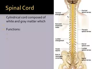
Ascending tracts Descending tracts Cell bodies Peripheralnervefibers Spinal Cord • Central gray nuclei and outer white matter (tracts)
PNS sensory motor CNS Peripheral and Central Nervous System Lesions
Spinal nerves & cord • 31 pairs with anterior and posterior root • C1 often doesn’t have posterior root • Spinal cord enlarged in C5 – T1 and L2 – S3 levels • Spinal cord ends at L1 vertebral level
Dermatomes & Myotomes • Dermatome – an area of skin innervated by a single posterior root • Myotome – a group of muscles innervated by a single anterior root
C4 T4 T10 L3 L5 Myotomes Dermatomes
Stretch Reflexes • Examples: • C5-6 = biceps tendon • C6 = brachioradialis • C7-8 = triceps tendon • L2-4 = patellar tendon • S1-2 = Achilles tendon
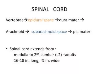
Conus medullaris Filum terminale Cauda equina Dura mater External Spinal Cord
Posterior Median Sulcus Posterior IntermediateSulci Lateral sulci Anterior Median Fissure External Structure – Spinal Cord
Posterior Funiculi Lateral Anterior Internal Structure – White Matter
Posterior Columns Lateral & Anterior Spinothalamic Tracts f. gracilis f. cuneatus Posterior & Anterior Spinocerebellar Tracts White Matter Organization:Ascending Pathways
Lateral Corticospinal Tract Rubrospinal Tract Reticulospinal Tracts Anterior Corticospinal Tract VestibulospinalTracts White Matter Organization:Descending Pathways
Lissauer’sTract Intersegmental Tracts White Matter Organization:Intersegmental Pathways
Posterior Columns Lateral & Anterior Spinothalamic Tracts L T A N L N White Matter Organization:Ascending Pathways
White Matter Organization:Descending Pathways Legs T A Neck
Posterior Motor Groups Intermediate Anterior Lateral Medial Grey Matter Organization
AKA Intermediolateral Cell Column Grey Matter Organization:Lateral Grey Horns: T1 – L2/3
Dorsomarginal nucleus Substantia gelatinosa Nucleus proprius Nucleus dorsalis (Clarke’s column) Grey Matter Organization:Posterior Cell Column
From Pritchard & Alloway: Fig. 3-7
Dorsomarginal n. Substantia gelatinosa Nucleus proprius Nucleus dorsalis (Clarke’s column) Motor nuclei Rexed Lamina