Download
1 / 51
520 likes | 1.42k Vues
Spinal Cord. Chapter 12 - continued . The Spinal Cord. The spinal cord extends from the foramen magnum to the level of the 1st or 2nd lumbar vertebrae It is enclosed within the vertebral column. The Spinal Cord. The spinal cord is a provides a two way conduction pathway to and from the brain

E N D
Spinal Cord Chapter 12 - continued
The Spinal Cord • The spinal cord extends from the foramen magnum to the level of the 1st or 2nd lumbar vertebrae • It is enclosed within the vertebral column
The Spinal Cord • The spinal cord is a provides a two way conduction pathway to and from the brain • It is a major reflex center
The Spinal Cord • The spinal cord is protected by bone, cerebro- spinal fluid, and meninges • Dura mater, arachnoid, pia mater
The Spinal Cord • Between the bony vertebrae and the dural sheath is a large epidural space filled with a soft padding of fat and a network of veins • Cerebrospinal fluid fills the subarachnoid space
The Spinal Cord • Inferiorly, the dural and subarachnoid membranes extend to the level of S2 while the spinal cord ends at L1 • Subarachnoid space beyond L1 is an ideal site for a spinal tap
The Spinal Cord • The spinal cord terminates in a tapering cone shaped structure called the conus medullaris
The Spinal Cord • A fiberous extension of the pia mater, the filum terminale extends inferiorly from the conus medullaris to attach to the posterior surface of the coccyx
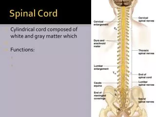
The Spinal Cord • There are 31 pairs of spinal nerves that arise from the cord by paired roots and exit from the vertebral column via the intervertebral formina • Each segment of the spinal cord is defined by a pair of spinal nerves that lie just superior to their corresponding vertebra
The Spinal Cord • The spinal cord has obvious enlargements where the nerves serving the upper and lower limb arise • Cervical enlargement • Lumbar enlargement
The Spinal Cord • Because the cord does not reach the end of the vertebral column, the lumbar and sacral spinal nerve roots angle sharply downward and travel inferiorly before reaching their intervertebral foramina • This collection of nerve roots at the inferior end of the vertebral canal is called the cauda equina • The arrangement reflects the fact that during vertebral column grows more rapidly than does the spinal cord
Embryonic Development • The spinal cord develops from the caudal portion of the embryonic neural tube • By the end of the 6th week each side of the developing cord has two clusters of neuroblasts that have migrated outwarded from the neural tube
Embryonic Development • The two clusters are the dorsal alar plate and a ventral basal plate • Alar plate neurons become interneurons • The basal plate neurons become motor neurons that sprout axons that grow out to the effector organs
Embryonic Development • Axons that emerge from alar plate cells form the external white matter of the cord by growing outward along the length of the CNS • The alar plates expand dorsally and the basal plates expand vertically to become the H-shaped mass of gray matter
Embryonic Development • Neural crest cells that come to lie alongside the cord form the dorsal root ganglia containing sensory nerve cell bodies, which send their axons to the dorsal aspect of the brain Neural crest cells
Cross Section of Spinal Cord • The spinal cord has two grooves that mark its surface • Anterior median fissure / Posterior medial sulcus
Gray Matter and Spinal Roots • These grooves run the length of the cord and partially divide it into right and left halves • Gray matter inside, the white matter outside
Gray Matter and Spinal Roots • The gray matter consists of a mixture of neuron cell bodies, their unmyelinated processes, and neuroglia (support cells)
Gray Matter and Spinal Roots • The white matter is composed of myelinated and unmyelinated nerve fibers that represent ascending, descending and transverse pathways
Gray Matter and Spinal Roots • The gray matter consists of mirror-image lateral gray masses connected by a cross-bar of gray matter called the gray commissure that encloses the central canal
Gray Matter and Spinal Roots • The two posterior projections of gray matter are the posterior (dorsal) horns; the anterior pair are the anterior (ventral) horns with lateral horns in the lumbar and thoracic portions of the cord
Gray Matter and Spinal Roots • The anterior horns house nerve cell bodies of the somatic motor neurons • These send their axons out via ventral roots of the spinal cord to the skeletal muscles
Gray Matter and Spinal Roots • The amount of ventral gray matter present at a given level of the spinal cord reflects the amount of skeletal muscle innervated at that particular level • Thus, the anterior horns are the largest in the areas where the innervation for limbs is present • Cervical enlargement / arms • Lumbar enlargement / legs
Gray Matter and Spinal Roots • The lateral horn neurons are autonomic (sympathetic) motor neurons that serve the visceral organs • Their axons also leave the cord via the ventral root
Gray Matter and Spinal Roots • Afferent fibers carrying impulses from peripheral sensory receptors form the dorsal roots of the spinal cord
Gray Matter and Spinal Roots • The cell bodies of the associated sensory neurons are found in an enlarged region of the dorsal root called the dorsal root ganglion or spinal ganglion
Gray Matter and Spinal Roots • After entering the cord, the axons take a number of routes • Some enter the posterior white matter of the cord or brain, others synapse with interneurons
Gray Matter and Spinal Roots • The dorsal and ventral roots are very short and fuse laterally to form the spinal nerves which are then considered part of the peripheral nervous system (PNS)
Gray Matter and Spinal Roots • The spinal gray matter can be divided further according to its neurons relative involvement in the innervation of the somatic and visceral regions of the body • The four zones are; somantic sensory (ss); visceral sensory (vs); visceral motor (vm); somatic motor (sm)
White Matter • The white matter of the spinal cord is composed of myelinated and unmyelinated nerve fibers that allow communication between different parts of the spinal cord and between the cord and the brain
White Matter • Nerve fibers run in three directions • Ascending / up to higher centers (sensory inputs) • Descending / down to the cord from the brain or from within the cord to lower levels (motor outputs) • Transversely / across from one side of the cord to the other (commissural fibers)
White Matter • The ascending and descending tracts make up most of the white matter of the spinal cord • Ascending tracts are shown in blue and labeled at left • Descending tracts are shown in red and labeled at right
White Matter • The white matter on each side of the column is divided into three white columns or funiculi and labeled according to their position (posterior, lateral, anterior) • Each funiculi contains several fiber tracts, and each tract is made up of axons with similar destinations and functions
White Matter • All major spinal tracts are actually part of multi- neuron pathways that connect the brain to the body • These ascending and descending pathways contain not only spinal cord neurons but also parts of peripheral neurons and neurons in the brain
White Matter • Generalizations about spinal pathways • Most pathways cross over from one side of the CNS to the other at some point • Most consist of a chain of two or three neurons that contribute to successive tracts • Most exhibit somatotopy, a precise spatial relationship among the tract fibers that reflects the orderly mapping of the body • All pathways and tracts are paired (right and left) with a member of the pair on each side of the spinal cord or brain
Ascending (Sensory) Tracts • The ascending pathways conduct sensory impulses upward, typically through chains of three successive neurons (first-, second, and third-order neurons) to various regions of the brain • Most of the incoming information results from stimulation of • General sensory receptors • Touch / pressure / temperature / pain • Stimulation of proprioceptors • Muscle stretch / tendon / joint
Ascending (Sensory) Tracts • In general, sensory information is conveyed along six main pathways on each side of the spinal cord • Four transmit impulses to the sensory cortex for conscious interpretation • Fasciculi cuneatus • Fasciculi gracilis • Lateral spinothalamic tract • Anterior spinothalamic tract • Two transmit impulses to the cerebellum to coordinate muscle activity • Anterior spinocerebellur tract • Posterior spinocerebellur tract
Ascending (Sensory) Tracts • Posterior funiculi (dorsal white column) • Fasciculi cuneatus • Fasciculi gracilis • Transmit information from the fine touch and pressure receptors and joint proprioceptors • These tracts comprize what is referred to as discriminative touch and conscious proprioception
Ascending (Sensory) Tracts • Lateral and anterior funiculi • Lateral spinothalamic tract • Anterior spinothalamic tract • Convey information on pain, temperature, deep pressure and course touch (undiscriminated)
Ascending (Sensory) Tracts • Anterior and posterior funiculi • Anterior spinocerebellar tract • Posterior spinocerebellar tract • Convey information from proprioceptors (muscle and tendon stretch) to the cerebellum which uses this information to coordinate skeletal muscle activity
Ascending (Sensory) Tracts • Since the spinocerebellar tracts do not terminate in the cortex, these pathways do not contribute to conscious sensation • The spinocerebellar tracts do not decussate and thus contribute to ipsilateral innervation
Descending (Motor) Tracts • The descending motor tracts that deliver impulses from the brain to the spinal cord are divided into two groups • Pyramidal tracts • All others
Descending (Motor) Tracts • Motor pathways involve two neurons, referred to as upper and lower motor neurons • The pyramidal cells of the motor cortex, as well as the neurons in subcortical motor nuclei that give rise to other descending motor pathways, are called upper motor neurons • The anterior horn motor neurons, which actually innervate the skeletal muscles are called lower motor neurons
Descending (Motor) Tracts • The lateral (pyramdial) and anterior corticospinal tracts are the major motor pathways concerned with voluntary movement, particularly precise or skilled movement
Descending (Motor) Tracts • The pyramdial tracts are also called the direct pathways because their axons descend without synapsing from the pyramidal cells of the primary motor cortex all the way to the spinal cord
Descending (Motor) Tracts • Pyramidal tracts synapse primarily with interneurons, but also directly with anterior horn motor neurons, principally those controlling limb muscles • The anterior horn motor neurons activate the skeletal muscles with which they are associated
Descending (Motor) Tracts • The remaining descending tracts include: • Rubrospinal • Anterior reticulospinal • Lateral reticulospinal • Vestibulospinal • Tectospinal
Descending (Motor) Tracts • The remaining tracts originate in different subcortical motor nuclei of the brain stem • These tracts were formerly lumped together as the extrapyramidal tracts • The current term is to label them indirect pathways or just the names of the individual pathway
Descending (Motor) Tracts • Although the cerebellum coordinates voluntary muscle activity, no motor efferents descend directly from the cerebellum to the spinal cord • The cerebellum influences motor activity by acting through relays on the motor cortex
Spinal Cord Trauma • Damage to the spinal cord is associated with some form of loss of function • Paralysis / loss of function • Paresthesis / sensory loss • Flaccid paralysis / motor loss • Spastic paralysis / upper motor neuron loss • Body regions below lesion • Quadriplegia / spinal cord injury - 4 limbs • Paraplegia / spinal cord injury - 2 limbs • Hemiplegia / brain injury - one side of body