Download
1 / 41
420 likes | 451 Vues
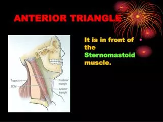
ANTERIOR TRIANGLE. SUB- MENTAL TRIANGLE Boundaries Contents. Lymph nodes are oval or bean-shaped structures found along lymphatic vessels that drain body parts. Normally, they are non-tender, soft and cannot be felt even though they are present.

E N D
SUB- MENTAL TRIANGLE Boundaries Contents
Lymph nodes are oval or bean-shaped structures found along lymphatic vessels that drain body parts. Normally, they are non-tender, soft and cannot be felt even though they are present. • This node is significant because it is the one node that is easy to discover the site of drainage. The lower lip, lower anterior gingivae, corners of the mouth, and skin and tissue of the chin drain to the submental node.
Most (70%) salivary gland tumors (SGTs) originate in the parotid gland. The remaining tumors arise in the submandibular gland (8%) and minor salivary glands (22%). Carcinoma of tongue
FLOOR • THYROHYOID • HYOGLOSSUS • MIDDLE CONSTRICTOR • INFERIOR CONSTRICTOR
Contents • Carotid sheath with its contents • Carotid arterial system • Internal jugular vein • Nerves of the carotid triangle • Glossopharyngeal nerve • Vagus nerve • Spinal accessory nerve • Hypoglossal nerve • Ansa cervicalis • Sympathetic chain • Apex of the Parotid gland • Deep cervical lymph nodes.
Deep fascia of the neck Pretracheal fascia Carotid sheath Investing layer Prevertebral fascia
Extent • Formation • Contents • Structures piercing the carotid sheath
Base of the skull to Tunica adventitia of aortic arch. • Anterior wall of the sheath is formed by pretracheal fascia and posterior wall by Prevertebral fascia. • Contents – varies, upper part has ICA, IJV AND LAST FOUR CRANIAL NERVES. Middle part has ICA, IJV AND VAGUS NERVE. Lower part has CCA, IJV AND VAGUS NERVE. • The roots of Ansa cervicalis are embedded in the anterior wall of the sheath
Tympanic branch • Pharyngeal branch • Branch to carotid body and sinus • Muscular branch to STYLOPHARYNGEUS • Tonsillar branches • Lingual branches
Meningeal branch • Superior laryngeal branch divides into • External laryngeal nerve • Internal laryngeal nerve • Auricular branch: • Cardiac branches • Pharyngeal branch • Right recurrent laryngeal nerve
Descendens hypoglossi • Branch to THYROHYOID
Ansa cervicalis • It is related to the anterior wall of the carotid sheath. • It consists of two roots, i.e. superior and inferior. Superior root (descendens hypoglossi) runs downward infront of carotid sheath, carrying the fibres of ‘C1’ and supplies the superior belly of omohyoid. • Inferior root (descendens cervicalis) runs downwards carrying the fibres of C2 and C3 and joins with superior root to form “ansa cervicalis”. • Ansa cervicalis supplies the sternohyoid, sternothyroid and inferior belly of omohyoid.
Drooping of the eyelid, a constricted pupil, absence of sweating, and a red flush to 1 side of the face caused by vasodilatation is indicative of Horner's syndrome caused by damage to the cervical sympathetic chain. • Horner's syndrome, in which the eyelid of the patient's left eye droops, and the pupil is contracted.