Download

1 / 28
470 likes | 2.15k Vues
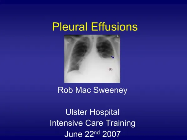
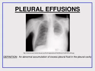
PLEURAL EFFUSIONS. http://intensivecare.hsnet.nsw.gov.au/five/images/pleural%20effusion%20CXR%202.jpg. DEFINITION - An abnormal accumulation of excess pleural fluid in the pleural cavity. ANATOMY AND PHYSIOLOGY IN A HEALTHY LUNG. The Right Lung -Makes up 56% of the total lung volume

E N D
PLEURAL EFFUSIONS http://intensivecare.hsnet.nsw.gov.au/five/images/pleural%20effusion%20CXR%202.jpg DEFINITION- An abnormal accumulation of excess pleural fluid in the pleural cavity
ANATOMY AND PHYSIOLOGY IN A HEALTHY LUNG The Right Lung -Makes up 56% of the total lung volume -Three lobes-the superior, middle and inferior, which are separated by the horizontal fissure and the oblique fissure. Horizontal fissure The Left Lung -Makes up 44% of the total lung volume -Two lobes which are separated by the oblique fissure. Oblique fissure Oblique fissure http://fitsweb.uchc.edu/ctanatomy/chest/review/segments-posterior.html
The main anatomy affected by pleural effusions are the layers in the Lung • There are two layers-the parietal pleura and the visceral pleura. • At the Hilum, the parietal pleura folds back on itself to become the visceral pleura. • The mesothelium is a membrane that forms the lining of the • pleura. • A simple squamous epithelium • The pleural fluid contains – • Glucose • Mesothelial cells • Macrophages • Lymphocytes • Sodium, potassium and calcium • Lactate Dehydrogenase http://www.nature.com/modpathol/journal/v18/n2/images/3800278f1.jpg
ANATOMY OF A HEALTHY LUNG ANATOMY OF A LUNG WITH A PLEURAL EFFUSION http://www.themesotheliomalibrary.com/pleural-effusions3.gif A pleural effusion is an accumulation of fluid between the parietal pleura and the visceral pleura.
ANATOMY OF A LUNG WITH A PLEURAL EFFUSION • The fluid accumulates due to the over production of pleural fluid by the mesothelial cells and separates the visceral and parietal pleura. • This fluid can not be drained by the lymphatic system, and so • therefore continues to accumulate, resulting in a pleural effusion. • The accumulation of fluid may also be due to changes in • hydrostatic pressure or oncotic pressure. • Other changes to anatomy include- • - Pleural thickening • - Mediastinal Pleural thickening • - Diaphragmatic Pleural thickening • - Presence of Pleural nodules • - Increase in pressure www.ncbi.nlm.nih.gov
There are 4 different fluids which can accumulate in the pleural space. • Blood HAEMOTHORAX • Pus EMPYEMA • Chyle CHYLOTHORAX • Serous fluid HYDROTHORAX • They can further be classified into TRANSUDATES and EXUDATES depending on • Chemical composition • Mechanism of fluid formation • Transudate due to systemic factors concerning the capillaries that supply blood to the thorax. • Exudates are the result of local factors such as inflammation of the plural or decreased lymphatic drainage.
Oncotic pressure Hydrostatic Pressure Increased peritoneal fluid
Transudates Congestive heart failure Cirrhosis of the Liver Nephrotic syndrome Pulmonary embolism Hypoalbuminemia Main causes:
What is an Exudate? Transudative and exudative pleural effusions are distinguished by measuring the lactate dehydrogenase (LDH) and protein levels in the pleural fluid. LDH is an enzyme which is important to the production of energy in cells. When cells die LDH is released and escapes into the blood. High levels of LDH indicate infection or cancer are present A cloudy, viscous fluid containing proteins and cellular debris which has escaped from blood vessels and has been deposited in tissues, or on tissue surfaces, usually as a result of inflammation. • Fluid extracted from pleura via thorococentesis as show in the image on this slide. http://medical-dictionary.thefreedictionary.com/thoracentesis
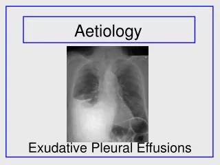
Causes of Exudative Pleural Effusions Parapneumonic causes Malignancy (carcinoma, lymphoma, mesothelioma) Pulmonary embolism Collagen-vascular conditions (rheumatoid arthritis, lupus) Tuberculous Asbestos exposure Trauma (haemathorax) Postcardiac injury syndrome Esophageal perforation Radiation pleuritis Drug use Once an exudative pleural effusion has been established from the fluid test, the causes can be investigated. An exudative pleural effusion is not a pathology in itself and is always caused by something else such as:
Mesothelioma • The Most common cause of mesothelioma is inhalation of asbestos fibres. • Asbestos fibres are very fine and sharp and once they have been inhaled they cannot be exhaled or coughed out http://www.asbestosclaimlawyermaryland.com/asbestosis-claim-asbestos-diseases
Mesothelioma http://www.mesotheliomacenter.org/about/pleural-mesothelioma.php Once asbestos fibres become deposited in the pleura they irritate and inflame the mesothelial cells affecting the adherens junctions allowing exudative fluid to flow into the pleural space.
Diagnosis of Pleural Effusions Medical history Physical examination Plain film chest x-ray – first line imaging CT Ultrasound MRI Diagnosing Pleural Effusions through Imaging
Meniscus shaped upper border Clear right side hemi-diaphragm and sharp costophrenic angle Area of homogenous Whiteness First line imaging – Chest x-ray Features on a PA radiograph http://intensivecare.hsnet.nsw.gov.au/five/images/pleural%20effusion%20CXR%202.jpg
These are approximations Berman, L., De Lacey, G and Raby, N. (2008) Accident and Emergency Radiology a survival guide, 2nd edition. Saunders, Elsevier. P262-263
Entire white-out of right hemi-thorax The hearthas been pushed towardsthe left side A large right side pleural effusion
Lateral decubitus chest radiograph Free layering pleural effusion emedicine.medscape.com/article/299959-media
Pleural Effusion Diagnosis through CT Imaging medical.siemens.com
Performing Chest CT Lung Window, showing anatomy of lungs Soft Tissue or Mediastinal Window http://emedicine.medscape.com/article/355524-media http://img.medscape.com/pi/emed/ckb/radiology/336139-362571-5581.jpg
Heart Right Lung Left Lung Crescent-shaped pleural effusion Ribs Aorta http://emedicine.medscape.com/article/35524/media
Loculated effusion (elliptical, pointed margins) in left major fissure http://emedicine.medscape.com/article/35524/media
Irregular soft-tissue thickening Mass, right upper lobe Pleural effusion Aorta http://emedicine.medscape.com/article/media
http://radiologyinthai.blogspot.com/2009_12_01_archive.html Axial CT images show a large mass (yellow stars) in the left lower lobe with a large left pleural effusion with focal pleural thickening (arrowheads). The lung mass can be seen better on the post thoracentesis image on the right.
Liver Ascites Spleen Diaphragm Pleural effusion Right Lung http://emedicine.medscape.com/article/35524/media
Advances in TechnologyPET/CT http://www.lung-cancer-blog.com/images/blogs/2-2008/pet-ct-scanner-41234692.jpg