Download
1 / 75
750 likes | 757 Vues
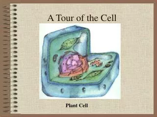
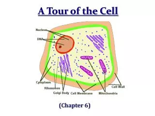
A Tour of the Cell. Anton Van Leeuwenhoek (1600’s) Credit for the first microscope Looked at pond water and saw “wee beasties”. Robert Hooke. Observed plant stems, wood, and cork (1600’s) Saw all the tiny chambers and called them CELLS What cell part did Hooke observe? Cell Wall.

E N D
Anton Van Leeuwenhoek (1600’s) • Credit for the first microscope • Looked at pond water and saw “wee beasties”
Robert Hooke • Observed plant stems, wood, and cork (1600’s) • Saw all the tiny chambers and called them CELLS • What cell part did Hooke observe? • Cell Wall
Robert Brown (1833) • Observed that cells had a dark structure within plant cells • Brown observed the nucleus and stated that all cells have nuclei (at this time no one knew that the nucleus has DNA)
Matthias Schleiden (1838) • Stated that all plants are made of Cells • Made many observations of plants around the area
Theodor Schwann(1839) • Stated that all animals are made of Cells • Observed many animal tissues
Rudolf Virchow(1855) • Stated that all cells come from pre-existing cells • Cells arise from the division of pre-existing cells
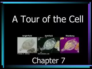
The Cell Theory Microscopes Provide the Windows to the World of the Cell
The Cell Theory • All living things are composed of cells • Cells are the basic unit of structure and function in living things • All cells come from pre-existing cells
The Size Range of Cells
Cell Sizes Average Animal Cell – 15 microns Average Plant Cell – 40 microns Average Eukaryotic Cell :10-100 microns Average Prokaryotic Cell: 1-10 microns
Geometric Relationships Explain Why Most Cells Are Microscopic
Cell or Plasma Membrane • “Fluid Mosaic” Model • Lipid Bilayer (made of phospholipids) • Proteins embedded throughout • Semi-permeable or Selectively Permeable
Cell Wall • provides support to the • perimeter of plant cells, some • protists, and bacterial cells
Nuclear Envelope/Membrane • Double Membrane that surrounds the nucleus • Lined with pores • Supported by nuclearlamina
Nucleolus • Inside the nucleus • Site of ribosome and rRNA synthesis
Endoplasmic Reticulum • (ER) • Rough ER • Intercellular transport of materials, particularly proteins; site where proteins leave ribosomes and are chemically modified
Smooth ER • breaks down toxic • substances, • regulates Ca levels, • synthesizes steroids and • other lipids
Golgi Apparatus • Modifies proteins and other substances from the ER for export from the cell
Lysosomes • Digest cellular waste • and foreign substances • Breakdown of lipids, • carbohydrates, and • proteins
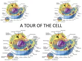
1 Nuclear envelope is connected to rough ER, which is also continuous with smooth ER Nucleus Rough ER 2 Membranes and proteins produced by the ER flow in the form of transport vesicles to the Golgi Smooth ER cis Golgi Nuclear envelop 3 Golgi pinches off transport Vesicles and other vesicles that give rise to lysosomes and Vacuoles Plasma membrane trans Golgi 4 5 6 Lysosome available for fusion with another vesicle for digestion Transport vesicle carries proteins to plasma membrane for secretion Plasma membrane expands by fusion of vesicles; proteins are secreted from cell • Relationships among organelles of the endomembrane system Figure 6.16
Peroxisomes • Contain an assortment of • enzymes that perform such • roles as detoxification of • alcohol, breaking down of • fatty acids • Produces H2O2 in theprocess
Plastids • May be called chromoplasts or leukoplasts • Store starch, fat or contain pigments such • as chlorophyll or carotenoids to capture energy from the sun
Mitochondrion Site of cellular respiration and synthesis of ATP, a source of chemical energy for the cell
Vacuoles • Store water, salts, • proteins, carbohydrates, • or enzymes
Cytoskeleton • Protein strands that give • the cell its shape and size • Helps organize the location • of organelles and their • activities
There are three main types of fibers the make up the cytoskeleton: 1) Microtubules 2) Microfilaments 3) Intermediate Filaments
Microtubules • Are made of the protein • tubulin • Shape and support the cell • Are responsible for the • separation of chromosomes • during cell division